Frequently Asked Questions — Mole Removal
Why do I need a dermatoscope examination for a simple mole?+
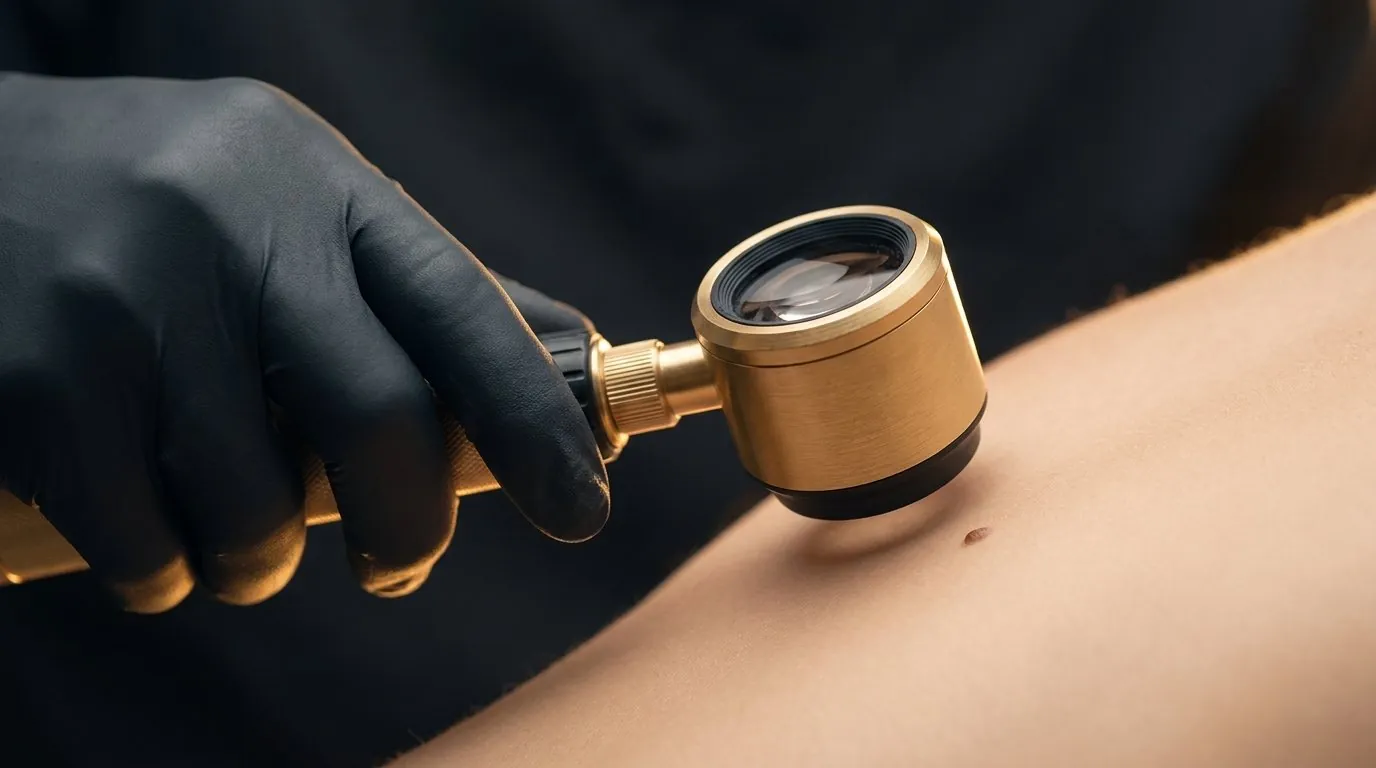
Because the surface tells only part of the story. Dermatoscopy shows the pigment network and vessel pattern beneath the surface, which is how a physician distinguishes an ordinary benign mole from a lesion that should be excised and examined instead of vaporised. It takes minutes, and it is the difference between removing a mole and removing information.
What happens if the scope shows something unusual?+
The plan changes. An atypical lesion is not lasered off — it is removed in a way that preserves the tissue for histological examination, or referred for specialist evaluation. This is precisely why scoping comes before removal, never after.
Which removal method will be used on my mole?+
That is decided at assessment. Small benign moles are commonly treated with mole removal laser techniques (CO2) or radiofrequency; protruding moles may suit shave removal; lesions needing histology are excised. Type, depth and location of the mole drive the choice.
Does mole removal hurt?+
The area is numbed with topical or injected local anaesthesia before removal, and comfort is checked before the mole is treated. Mild tenderness at the site afterwards is common, and aftercare is explained on the day.
Will there be a scar?+
Any removal leaves a mark that matures over weeks to months; the aim of matching method to mole is to keep that mark as inconspicuous as the lesion and location allow. Wound care instructions — and sun protection over the healing site — meaningfully influence the final result.
Can all moles be removed by laser?+
No — and that is the point of scoping first. Laser ablation is appropriate for moles with a clearly benign dermatoscopic pattern. A lesion with atypical features needs excision with histology, not vaporisation.
How long does the site take to heal?+
A small scab typically forms and separates over one to two weeks, with residual redness fading over the following weeks. Healing time varies with the mole's size, depth, the method used and the site on the body.
Will the mole grow back?+
A completely removed mole does not usually recur, though deeper dermal components can occasionally re-pigment and may need a touch-up assessment. Any regrowth should itself be re-scoped rather than assumed harmless.
Should I use an over-the-counter mole removal pen?+
No. No matter what a mole removal cream Singapore retailer promises, creams, corrosive pens and salon cautery all remove tissue without assessment, frequently scar, and destroy the possibility of histological examination. A mole should be assessed by a physician with a dermatoscope before anyone removes it — that is the standing rule of this practice.
How many moles can be removed in one session?+
Multiple scoped, benign moles can commonly be treated in a single sitting. The number is agreed at consultation based on their sites and the aftercare involved.
How much does mole removal cost in Singapore?+
The mole removal price in Singapore depends on the removal method, and the number, size and location of the moles being treated — so the mole removal Singapore cost for your case is quoted individually after the dermatoscopic assessment at consultation. Fees are always confirmed with you before any procedure is done.
Is there such a thing as scarless mole removal?+
Truly 'scarless' mole removal is a marketing phrase, not a medical one — any removal that reaches the mole leaves some mark. What careful technique can do is keep that mark small, flat and inconspicuous by matching the method to the mole's type, depth and location, and by protecting the healing site afterwards.
Is flat mole removal different from raised mole removal?+
Yes. A flat mole sits at or near the junction of the skin layers and is usually removed by CO2 laser in fine, controlled layers, while a raised dermal mole extends deeper and may respond better to radiofrequency or shave removal. This is one more decision the dermatoscope settles before treatment begins.
Can laser be used for wart removal?+
Laser ablation is one of the methods doctors use for viral warts that have not responded to simpler treatment, but it is not usually the first step. Because the virus can persist in the surrounding skin, warts can recur after any method. Whether a growth is a viral wart, an age wart or a mole is established first, since each is managed differently.
Is an age wart the same as a viral wart?+
No. An age wart is a seborrhoeic keratosis: a common, non-viral growth that becomes more frequent from middle age and is not contagious. A viral wart is caused by the human papillomavirus and can spread. Both can be mistaken for a mole, which is why a lesion is examined with a dermatoscope before removal is planned.
Should a mole that itches or bleeds be checked?+
Yes. Itching, bleeding or crusting without an obvious injury are changes that count under E for Evolving in the ABCDE screen. Many turn out to be irritation from clothing, a bag strap or shaving, but the mole should be examined with a dermatoscope before it is either removed or left alone, rather than treated at home.
Can I have a mole removed for cosmetic or face-reading reasons?+
Yes. A benign mole can be removed simply because you would prefer it gone, whatever the reason. The pathway is the same: the mole is examined under the dermatoscope first, the method is matched to its type and site, and any mole with atypical features goes for histology instead of cosmetic removal. Some mark is left wherever a mole is removed, which is worth weighing on the face.
Who is not a good candidate for cosmetic mole removal?+
A mole with atypical features on dermatoscopy, or one that is changing, bleeding or unlike your other moles, is excised for histology or referred instead of being removed cosmetically. A keloid tendency, blood-thinning medicines, slow healing, local anaesthetic allergy, infection at the site and pregnancy each change the timing or the method. This is decided at consultation.
Is mole removal worth it?+
It is worth considering when a mole has a benign pattern on dermatoscopy and bothers you cosmetically or physically, and you accept that a mark will remain. It is not suited to moles with atypical features, which need histology or referral, or to anyone expecting a mark-free result. Leaving a benign mole alone is also a valid choice.
How long does mole removal last?+
A removed benign mole may not return, but this depends on the mole's depth, the method used and how the site heals. If deeper mole cells remain, pigment can reappear, and new moles can still form elsewhere. Review timing is set at consultation, and any regrowth is re-examined rather than simply treated again.
What are the disadvantages of mole removal?+
Any removal leaves a mark that matures gradually, and it can occasionally be depressed, raised, lighter or darker. Healing needs wound care and sun protection, and infection, bleeding or keloid scarring are uncommon risks. Laser and radiofrequency limit tissue available for histology, so atypical moles are excised instead. Regrowth is possible.