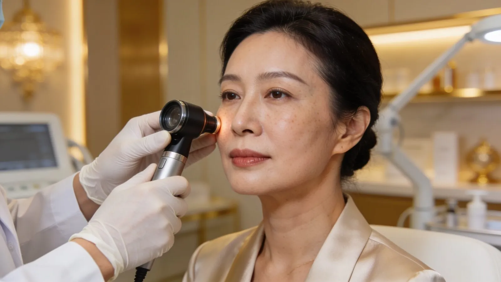
Mole removal in Singapore starts with one question: has this mole been properly examined, with dermatoscopy where indicated, to tell a benign mole from one that is not? Only then is a method chosen, such as shave removal, radiofrequency or laser ablation, or elliptical excision with histology, matched to the mole's depth and the need for diagnosis.
Key facts
- ABCDE assessment
- Asymmetry, Border irregularity, Colour variation, Diameter and Evolution — the screening framework for pigmented lesions since 1985
- Junctional, compound, dermal
- The three depths at which naevus cells sit; depth influences both appearance and the removal method that clears the lesion
- Shave removal
- Tangential removal at skin level under local anaesthesia; suits raised dermal moles
- Radiofrequency / laser ablation
- Layer-by-layer vaporisation of small superficial moles; no sutures
- Elliptical excision
- Full-thickness removal with sutures; the method when complete depth and histology are the priority
- Histology
- Microscopic examination of removed tissue — the only way to confirm what a lesion actually was
- Local anaesthesia
- All methods are performed awake under local anaesthetic infiltration
The assessment comes first — always
A mole is a colony of pigment cells, and the entire point of a medical assessment is to establish which kind of colony you are dealing with before it is destroyed. The ABCDE framework — asymmetry, border, colour, diameter, evolution — has guided this screening since Friedman and colleagues formalised it in 1985, and dermatoscopy lets a trained eye read the pigment pattern beneath the surface. Most moles are innocent. The assessment exists for the minority that are not, because the worst possible outcome in this field is an atypical lesion vaporised by a machine with nothing left to examine.
This is why I am cautious about the walk-in, point-at-it, burn-it-off model of mole removal. A doctor should be able to say why a mole looks benign — not just that it does. Any mole that has changed in size, shape or colour, bleeds, itches, has irregular borders or mixed colours, or simply looks different from its neighbours earns a closer look, and sometimes excision with histology rather than ablation.
“The dangerous question is not how to remove a mole. It is whether anyone competent looked at it first.”
Dr Sin YongOn why assessment precedes every removal
Why moles are removed
Three honest reasons. Medical suspicion — the mole needs to come off completely and go to the laboratory. Friction and function — a raised mole that catches on razors, spectacles, collars or combs. And appearance — a prominent facial mole the owner has simply never liked. All three are legitimate; they just lead to different methods, and pretending one method fits all is how people end up with the wrong scar, a regrown mole, or a missed diagnosis.
Shave, ablate or excise — how the method is chosen
A raised, clearly benign dermal mole often suits shave removal: the protruding portion is taken flush with the skin under local anaesthetic, healing over a week or two. Small, flat, superficial moles can be ablated by radiofrequency or laser in controlled layers, which needs no sutures and suits multiple small lesions in one sitting. A mole where depth matters — anything atypical, anything the assessment flags — is excised as an ellipse with a fine suture line, because excision is the only method that removes the full depth and produces a specimen for histology.
Each method trades something. Shave and ablation leave the base of the naevus behind, so a proportion of moles partially re-pigment over the years — not a complication, but a known behaviour worth hearing about before you choose. Excision removes everything but leaves a line rather than a dot. Matching the trade-off to the mole and its location is the actual skill.
What determines the scar
The scar is decided by four things: the removal depth, the location, your own healing tendency, and aftercare. Central-face skin heals remarkably well from superficial work; the jawline, chest and shoulders scar more assertively, and anyone with a keloid history needs that factored in before any skin surgery, however minor. Sun protection on the healing site matters in Singapore's ultraviolet climate, because a healing wound that tans becomes a mark that lingers. A realistic framing: removal exchanges a mole for as subtle a scar as the site allows — usually subtle, never literally nothing.
Aftercare, and what regrowth means
Ablated and shaved sites crust and re-epithelialise over one to two weeks; excisions have sutures removed at about a week on the face. A scab picked early is a scar invited, so the instruction sheet is short but non-negotiable: keep it moist, keep it clean, keep sun off it. If pigment reappears at a shaved or ablated site months later, it is usually residual naevus cells at the base doing what naevus cells do — but any regrowth is reassessed rather than casually re-burned, because regrowth in a previously removed lesion deserves eyes on it.
Singapore specifics worth knowing
Mole removal here is a medical procedure performed by doctors, not a beauty-salon service — and pigmented lesions in Asian skin deserve particular respect, because darker skin types both pigment more readily after inflammation and can develop melanomas in less-watched sites such as palms, soles and nails. The practical message: choose assessment-led removal, and use the appointment to have the rest of your skin glanced over while you are there. It takes minutes and occasionally matters enormously.