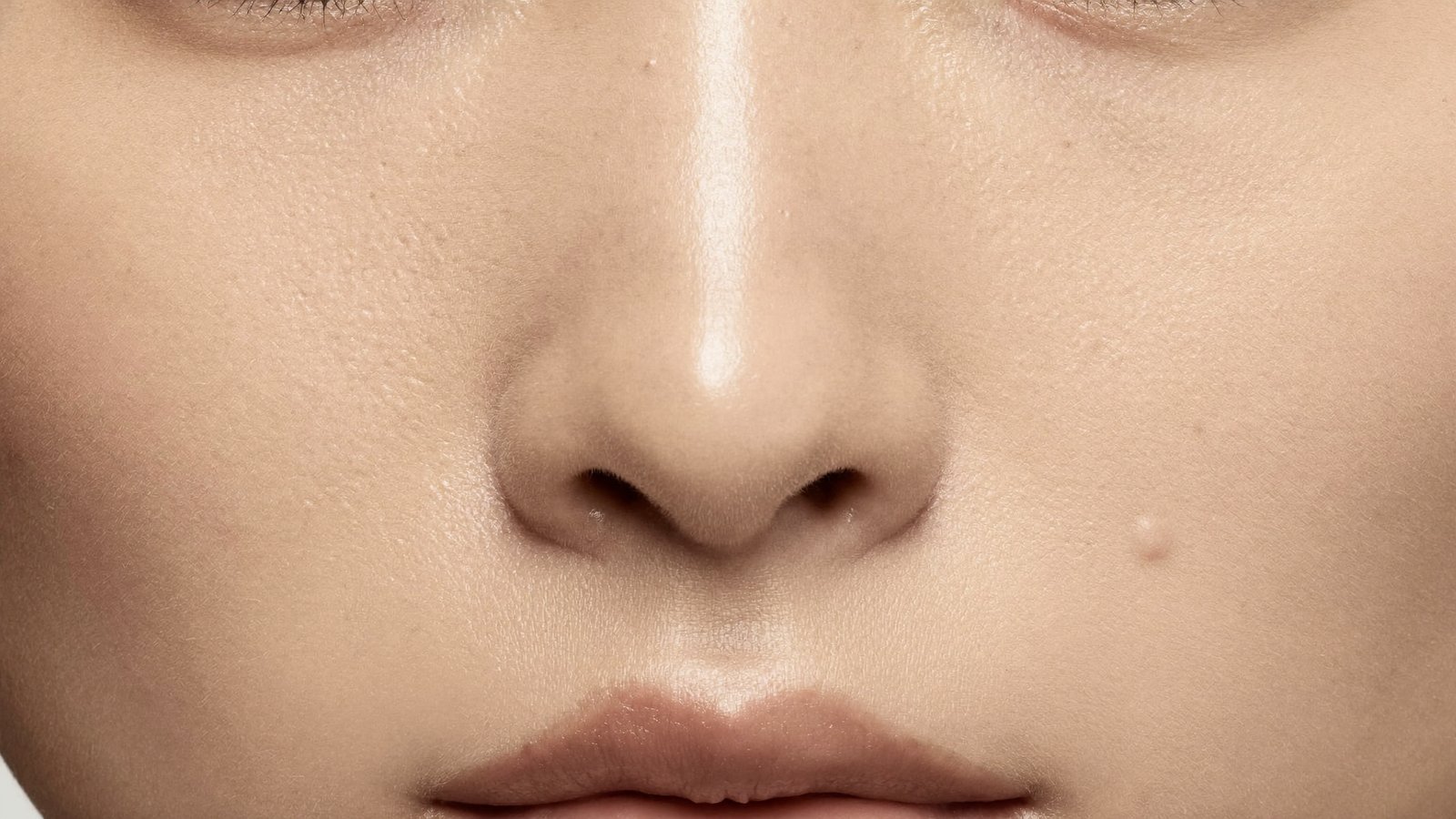
A cherry angioma, the spot many people call a red mole, is a small, benign cluster of blood vessels in the skin, not a pigment mole. These bright red to purple dots become more common with age and do not turn into melanoma. Removal is optional and cosmetic; at this clinic it is done with R2 Glow, but a red spot that is new, bleeding or unlike the others is examined first.
Red mole or something else? The red spots compared
Most spots people call a red mole are cherry angiomas. A few red spots are something else, and that is the reason a red mole is examined before it is removed. The comparison below sets out the red spots that are confused with each other, what each usually looks like, and what normally happens next. It is a guide to the conversation, not a way to diagnose yourself.
Red spot
What it usually looks like
What it is
What happens next
Cherry angioma
Bright red to purple dot or smooth dome, often on the trunk and upper arms
A benign cluster of capillaries; no pigment cells
Left alone, or removed for cosmetic reasons after examination
Spider angioma
A red centre with fine vessels radiating from it, blanching on pressure
A red lump that appeared quickly and bleeds easily
A fast-growing vascular lesion
Examined promptly, not treated as an angioma
Angiokeratoma
Dark red to black with a rough surface, often on the legs
A vascular lesion with surface thickening
Examined with a dermatoscope
Red or pink skin cancer
Rare; pink or red with little pigment, new or growing
The reason any changing red spot is examined first
Removed for histology or referred, never cauterised unexamined
A red mole on the face, cheek or body that has been there for years, matches your other spots and has not changed is usually a cherry angioma. A spot that is new and growing, bleeds without a knock, looks different from the others or crusts and breaks down is examined under a dermatoscope before anything is decided. The same rule applies to brown spots on mole removal, and to the waxy stuck-on growths described under seborrhoeic keratosis.
This topic is part of the main guide to mole removal in Singapore, which covers what it treats, who it suits, the risks and how Dr Sin Yong approaches it.
Illustrative. A red spot is examined before any decision about removal is made.
Your assessment
What happens at your assessment
Dr Sin Yong examines you himselfHe looks at and feels the area, and goes through what was done and when.
You hear what he findsHe explains what has happened in the skin and tissue, in plain words.
You decideHe sets out the options: treat, monitor or leave alone. Nothing is done on the day unless you choose it.
Who you will see: Dr Sin Yong. He performs every assessment and treatment himself.
A cherry angioma is a small, benign cluster of blood vessels in the skin, not a mole, seen as a bright red to purple dot that becomes more common with age. It contains no pigment cells and does not turn into melanoma. Removal is optional and cosmetic, but a new, bleeding or unusual red spot is examined first.
Key facts
What it is
A benign capillary growth: a cluster of small blood vessels, not pigment cells
Other names
Campbell de Morgan spot, senile angioma, cherry haemangioma
Typical look
Bright red to purple, flat or dome-shaped, from a pinpoint to a few millimetres
Who gets them
Increasingly common with age; reviews estimate around three-quarters of people over 75 have them
Malignant potential
None, but a small number of red lesions are something else, so diagnosis comes first
Removal options
At this clinic, R2 Glow for flat and raised angiomas, after examination; other clinics also use electrocautery, radiofrequency or cryotherapy
First step
Examination, with a dermatoscope where indicated, before anything is removed
Cherry angiomas are the red moles many people collect with age, and they are harmless vascular spots rather than pigment moles. The practical question is diagnosis, not danger: a dermatoscope separates them from the few red lesions that are something else. If a spot is confirmed and bothers you, the removal method is matched to its size and site.
What is a cherry angioma, and why is it called a red mole?
A cherry angioma is a benign growth of tiny blood vessels in the upper layers of the skin. It looks like a mole because it is a small, well-defined dot, and many people call it a red mole for that reason. Biologically it is a different thing: a mole is a cluster of pigment-producing cells, while a cherry angioma is a cluster of capillaries, and its colour comes from the blood inside them rather than from melanin.
Dermatologists know the same lesion as a Campbell de Morgan spot, a senile angioma or a cherry haemangioma. They range from a pinpoint red fleck to a smooth dome a few millimetres across, bright red when young and often darker, towards purple, as they mature. Most sit on the trunk, upper arms and shoulders, and they are unusual on the hands and feet.
Their importance is not medical. They cause no symptoms and carry no risk of turning into cancer. But they are visible, they can bleed when nicked by a razor or caught on a strap, and they are easy to confuse with other spots that do matter, which is the real reason to understand them.
Cherry angiomas: small bright red dome-shaped spots. Illustrative video, not a patient.
Why do cherry angiomas appear as we get older?
Age is the clearest driver. Cherry angiomas become noticeably more numerous from around the age of 40, and reviews estimate that around three-quarters of people over 75 have them. A smaller proportion of people notice their first ones in their twenties, or even in adolescence. New spots appearing gradually over the years is the expected pattern, not a sign that something has gone wrong.
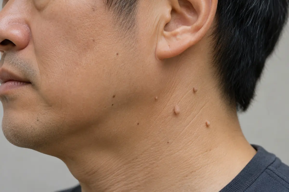
Skin tags are harmless and are removed for comfort or appearance. Illustrative image, not a patient.
Molecular studies have found specific acquired mutations, in genes such as GNAQ and GNA11, in the vessel cells of many cherry angiomas. That helps explain why they behave as small, stable growths rather than as a rash or inflammation, and why no cream, diet or supplement makes them recede. A gradual accumulation reflects how skin vessels behave over time, not anything you did or failed to do with your skincare.
Other associations are described but are far less common: pregnancy, certain medicines and chemical exposures, and, rarely, a sudden eruption of many new angiomas alongside other illness. A slow accumulation of a few spots with age needs no investigation. A sudden crop of many new lesions, or angiomas appearing while you feel generally unwell, is worth mentioning to a doctor.
“Do not remove your mole until you have scoped it.”
Dr Sin YongOn examining any spot before it is removed
Sudden red moles: what does it mean when many appear at once?
Sudden red moles are usually new cherry angiomas, and noticing several at once is common. Often the spots have built up gradually and are simply noticed for the first time, after a haircut, a holiday photo or a new mirror. A true eruption, with many new red spots in a short period, is less common and is worth showing to a doctor, because reviews have linked cherry angiomas with certain medicines, chemical exposures and changes in the immune system.
What can bring on a crop of red moles
Age, particularly from around 40, which explains most new spots.
Pregnancy and other hormonal change, when several kinds of vascular spot become more common.
Some medicines and chemical exposures, described in case reports and reviews.
A weakened immune system or a general illness, where the spots are one clue among others.
Red dots that are not cherry angiomas
The lookalike that matters most for sudden red dots is petechiae: flat, pinpoint red or purple spots caused by tiny bleeds into the skin. A cherry angioma usually fades at least partly when a clear glass is pressed firmly over it; petechiae do not fade, and they often appear in a cluster, sometimes with bruises. Spider angiomas have a red centre with fine vessels running out from it. Under a dermatoscope a cherry angioma shows well-defined red to purple vascular compartments, which settles most cases at the first visit.
What you can do at home, and what you cannot
You can photograph new spots beside a ruler, date the photographs and compare them later. You can press a clear glass over a new red dot to see whether it fades. You cannot tell an atypical red lesion from a harmless one at home, and cutting, freezing or burning spots with home kits removes the chance to check what they were. In Asian skin, picking and home removal can also leave a brown mark that outlasts the spot itself.
When it needs a doctor
Seek medical care the same day if new red or purple dots do not fade under a glass, especially with bruising, bleeding gums, nosebleeds, fever or feeling unwell. Many new angiomas within a short time, or spots appearing alongside weight loss or another illness, are worth raising with your GP. Once a spot is confirmed as a cherry angioma, it can be left alone or removed for cosmetic reasons with R2 Glow; brown or changing spots are assessed under mole removal.
Cherry angioma, mole or seborrhoeic keratosis: how do you tell them apart?
Colour and texture are the first clues. A cherry angioma is red to purple and smooth. A mole is made of pigment cells, usually tan, brown or black, flat or raised, and it can change in ways that matter. A seborrhoeic keratosis, the so-called age wart, is a waxy, stuck-on growth, tan to dark brown, that looks as though it could be lifted off with a fingernail. Skin tags, soft flesh-coloured flaps, often sit on the same skin. All of these become more common with age, which is why people confuse them.
Older angiomas can darken towards purple or look almost black, and that is where self-assessment fails. A dark, dome-shaped spot could be an angioma that has clotted, a pigmented seborrhoeic keratosis or a pigmented lesion that needs attention, and from the surface they can look alike. Under a dermatoscope the distinction is usually much clearer: angiomas show well-defined red to purple vascular compartments, while moles show a pigment network.
Other red spots belong in the comparison too. Facial thread veins are fine lines of dilated vessels rather than a dot. Spider angiomas have a red centre with fine vessels radiating from it, and several appearing together are worth mentioning to a doctor because they have different associations, including pregnancy and liver disease. A red lump that appeared quickly and bleeds easily may be a pyogenic granuloma, and rarely a skin cancer can be pink or red with little visible pigment.
When should a red spot be checked by a doctor?
A red spot should be checked when it is new and growing quickly, bleeds without an obvious injury or keeps re-bleeding, crusts or breaks down, looks different from your other angiomas, or sits somewhere unusual. Simply being unsure is also a good reason. None of these mean the lesion is serious; they mean the decision should rest on an examination rather than an assumption. A dated phone photograph of the spot beside a ruler is a useful record if you are unsure whether it is genuinely changing.
Examination is quick. The spot is looked at under good light and, where indicated, with a dermatoscope, which shows the vessel pattern beneath the surface. A classic cherry angioma can then be left alone or removed for cosmetic reasons. Anything atypical is not cauterised away: it is removed in a way that preserves tissue for histology, or referred, as with any lesion that does not look straightforward. The same principle runs through mole removal at the practice.
It is worth having the rest of your skin glanced over at the same visit. People with many angiomas often have moles and seborrhoeic keratoses as well, and the appointment that settles one red spot can settle several brown ones. The questions worth asking before any lesion is removed are set out in the mole removal guide.
Cherry angioma removal: am I suitable, and which method fits the spot?
You are usually suitable for cherry angioma removal if a dermatoscope confirms the spot is a cherry angioma and it bothers you because of how it looks, or because it catches on clothing or a razor. Removal is optional. A red spot that is growing quickly, bleeds without injury or looks different from your other spots is examined first and not treated as an angioma until that question is settled.
Light absorbed by haemoglobin heats and closes the vessels
Flat and raised angiomas after examination, with settings matched to the size and height of each spot
Replace examination, or histology for a spot that looks atypical
Electrocautery or radiofrequency (used at other clinics)
A fine energy tip seals and removes the vessels under local anaesthetic
Raised angiomas that sit above the skin
Avoid a small mark where a larger angioma was
Cryotherapy
Freezing destroys the small vessels
Some small lesions, in some clinicians’ hands
Rule out a pale or dark mark afterwards
Home pens, freezing kits or tying off
Corrosion, freezing or cutting off the blood supply without examination
Not advised
Check what the spot was, or stop it bleeding
What decides the method is the size of the angioma, how raised it is, where it sits and your skin type. On the face, where a mark matters more, the plan is more cautious. In Asian skin a treated site can darken for a while or, occasionally, leave a pale spot, so sun protection while it heals is part of the treatment, not an extra.
Who should wait: anyone with a spot that has changed recently until it has been examined, and anyone with broken or inflamed skin over the area until it settles. A fresh tan is also a reason to delay laser. Removal treats the angiomas present, not the tendency to form them, so new ones may appear later. For other red and brown spots, see mole removal.
How are cherry angiomas removed, and what are the trade-offs?
Removal is optional and cosmetic. At this clinic Dr Sin Yong uses R2 Glow for cherry angiomas, after examination. Other clinics also use electrocautery or radiofrequency, which seal and remove the vessels with a fine tip under local anaesthetic; vascular laser, in which light absorbed by haemoglobin heats and closes the vessels; and cryotherapy, which some clinicians use for small lesions. Which suits a given angioma depends on its size, how raised it is, where it sits and your skin type, and the options are discussed at assessment rather than chosen from a menu.
Each method trades something. At this clinic both flat and raised angiomas are treated with R2 Glow, with settings matched to the size and height of each spot. A small mark can remain where a larger angioma has been treated, and in Asian skin a treated site can darken for a while or, occasionally, leave a pale spot, so sun protection while it heals matters. A randomised trial comparing cryotherapy and electrosurgery for common benign lesions, including cherry angiomas, recorded pigment change and occasional small depressed marks in a minority of treated lesions.
Removal treats the lesions present, not the tendency to form them. New angiomas may continue to appear over the years, and some people return to have new ones dealt with as they arise. Home methods such as corrosive pens, freezing kits, tying off or picking are not advised: angiomas bleed readily, and destroying a lesion that has not been examined removes the chance to check what it was.
What happens at the visit, and what affects the cost?
The visit starts with the examination described above, followed by a discussion of which lesions to treat and how. Treated sites are numbed with local anaesthetic or a topical cream, and treatment of each small lesion is brief. A small crust may form and separate as the skin heals. Aftercare is simple: keep the area clean, do not pick, and protect it from the sun. A treated site that bleeds repeatedly, becomes increasingly painful, red and warm, or is not healing as expected should be reviewed rather than watched at home.
Cost depends on how many lesions are treated, their size and site, and the method used. Singapore's healthcare advertising rules do not allow prices for licensable healthcare services to be published, so fees are set out at consultation, after the spots have been examined and before anything is done.
Dr Sin Yong, MBBS (NUS), MRCS (Edinburgh), MSc Aesthetic Medicine with Distinction (Queen Mary, London), MSc Practical Dermatology (Cardiff), examines skin lesions personally on Orchard Road, and will say plainly when a red spot can simply be left alone. Where removal is suitable, it is carried out at the clinic after that assessment.
Key takeaways
A cherry angioma is a benign cluster of tiny blood vessels, not a mole; it causes no symptoms and does not turn into cancer.
Cherry angiomas become more numerous with age, especially from around 40, and no cream, diet or supplement makes them recede.
Red or purple dots that do not fade under a pressed glass, especially with bruising, bleeding gums or fever, need medical care the same day.
A red spot that is new and growing, bleeds without injury or looks different from your others is examined under a dermatoscope first, and anything atypical is removed for histology or referred.
Removal is optional and cosmetic; at this clinic Dr Sin Yong uses R2 Glow after examination, and sun protection while the site heals is part of the treatment.
Removal treats the angiomas present, not the tendency to form them, so new ones may appear later.
Related Related: sebaceous hyperplasia — the yellow bumps on oily skin, their lookalikes, and how each is flattened.
When a red spot turns out to be a pigmented mole, what decides the mole removal price is set out factor by factor.
What a 577 nm yellow laser targets, and what is used for red spots here instead, is set out under yellow laser benefits.
Frequently Asked Questions
A small, bright-red dot on the cheek is often a cherry angioma, a harmless cluster of tiny blood vessels rather than a pigment mole, although they are more common on the trunk and upper arms. Other red spots can look similar, so a spot that is new and growing quickly, bleeds without injury or looks different is examined with a dermatoscope before anything is done.
A cherry angioma is a small, harmless growth of tiny blood vessels in the skin, seen as a bright red to purple dot or smooth dome. It is also called a Campbell de Morgan spot or senile angioma. Cherry angiomas become more common with age, most often on the trunk and arms, contain no pigment cells and do not turn into cancer. A new, bleeding or unusual red spot is examined before anything is done.
Red mole is an everyday name for a small red spot on the skin, and in most adults it is a cherry angioma, a cluster of tiny blood vessels rather than a true mole made of pigment cells. The same name is sometimes used for spider angiomas, pyogenic granulomas or, rarely, a pink skin cancer, which is why a red mole that is new, growing or bleeding is examined with a dermatoscope first.
Age is the main cause: cherry angiomas become steadily more common from around 40. Acquired changes in genes such as GNAQ and GNA11 have been found in the vessel cells of many angiomas. Pregnancy, some medicines, chemical exposures and changes in the immune system have also been linked with them. Diet and skincare do not cause them, and a sudden crop of many new spots is worth showing to a doctor.
Usually not. Once a cherry angioma has formed it tends to stay, and new ones often appear with age. They are harmless, so leaving them alone is a reasonable choice. A spot that shrinks, bleeds or changes noticeably should be examined, because a change is the cue for a doctor to look rather than a sign it is clearing.
Treated sites are numbed with a topical cream or local anaesthetic first, and treatment of each small spot is brief. Comfort is checked as each spot is treated. Afterwards a small crust may form and separate as the skin heals; keep the area clean, do not pick it, and protect it from the sun.
Cherry angiomas are not a sign of liver disease; they are common with age. A different red spot, the spider angioma, has a red centre with fine vessels radiating outwards, and several of these together are worth mentioning to a doctor. A dermatoscope tells the two apart.
Yes, after examination. On the face the method and settings are chosen with more caution, because a small mark or a temporary change in colour is more visible there. In Asian skin a treated site can darken for a while, so sun protection while it heals is part of the plan.
Usually that you have a cherry angioma, a harmless cluster of small blood vessels that becomes more common with age. It is called a red mole because it looks like a small dot, but it contains no pigment cells and does not turn into melanoma. A red spot that is new and growing, bleeds without injury or looks unlike the others should be examined.
New cherry angiomas appearing gradually from around the age of 40 is the expected pattern. A red spot is worth examining if it grows quickly, bleeds without a knock, crusts, looks different from your other spots or appears in a sudden crop. Examination with a dermatoscope usually settles the question quickly, and most red spots can then be left alone.
Yes. At this clinic Dr Sin Yong treats cherry angiomas, flat or raised, with R2 Glow, whose light is absorbed by blood in the small vessels. Each spot is examined first and the settings are matched to its size and site, and anything atypical is removed in a way that keeps tissue for histology.
No. Cherry angiomas are benign growths of small blood vessels and do not turn into cancer. The reason to have a red spot examined is to confirm that it is a cherry angioma, because a small number of red lesions are something else.
Usually, yes. Most spots people call red moles are cherry angiomas, which are vascular growths rather than true moles. A true mole is made of pigment cells and is usually brown or black, so the two are different lesions with different assessments.
Cherry angiomas become more numerous with age, particularly from around 40, so new ones appearing over the years is the expected pattern. A sudden crop of many new lesions, or lesions appearing while you feel unwell, is worth mentioning to a doctor.
Yes. Because they are made of blood vessels, they can bleed when nicked by a razor, scratched or caught on clothing, and firm pressure usually stops it. A spot that bleeds without injury or keeps re-bleeding should be examined.
It is not advised. Corrosive pens, freezing kits and picking remove tissue without an examination, can bleed and scar, and leave nothing to check if the lesion was not what it seemed.
New angiomas can continue to appear elsewhere over the years, because removal treats the lesions present rather than the tendency to form them. If a treated spot re-forms, it is re-examined rather than simply treated again.
It can. Any removal leaves some mark, and its size depends on the angioma, the method and your healing. Larger or raised lesions are more likely to leave a small flat or pale spot, and sun protection while healing helps limit darkening in Asian skin.
Prices are not published on this site. Cost depends on the number, size and site of the angiomas and the method used, and is set out at consultation after examination.
Evaluation and Comparison of the Efficacy and Safety of Cryotherapy and Electrosurgery in the Treatment of Sebaceous Hyperplasia, Seborrheic Keratosis, Cherry Angioma, and Skin Tag: A Blinded Randomized Clinical Trial Study. Health Science Reports, 2024. source
Clinical Relevance of Cherry Angiomas. Actas Dermo-Sifiliográficas, 2023. pubmed.ncbi.nlm.nih.gov
Diagnosing Common Benign Skin Tumors. American Family Physician, 2024. pubmed.ncbi.nlm.nih.gov