Published 26 October 2026 · Reviewed by Dr Sin Yong
Heavy or pillowy cheeks, puffiness below the eyes, filler that has moved, lumps: cheek filler can go wrong months or years after the injection, and some people are then told it can only be cut out. That leaves a scar, can leave a dent and can injure the nerves that move your smile. Dr Sin Yong maps the filler with ultrasound and treats it without an incision. Get assessed first.
Short answerGet assessed before you agree to surgery. Cheek filler usually goes wrong in a few ways: too much filler, giving ‘chipmunk’ cheeks or a ‘pillow’ face (Lim 2024); filler that has moved towards the under-eye or the smile lines; puffiness on the upper cheek, called malar oedema (Funt 2011); lumps; or a delayed reaction. Ultrasound shows which, and what the filler is likely made of. Hyaluronic acid is dissolved after a skin test. Dr Sin Yong treats Sculptra, Radiesse, Ellansé and silicone with the Sculpting mode of Time Freeze Laser LCLR®, without an incision.
Get assessed. Cutting filler out of the cheek leaves a scar, can leave a dent, can injure the nerves that move your smile or give the cheek feeling, and cannot be undone.
Non-absorbable filler
In 325 people with complications from non-absorbable filler, the cheeks were the commonest site (66.6%), and problems most often began 1 to 5 years after injection (Mortada 2022).
Collagen stimulators
In 130 people treated with poly-L-lactic acid (Sculptra), nodules were the commonest side effect (8.5%), mostly felt rather than seen, and most frequent in the hands and cheeks (Palm 2010).
Filler may expand
On MRI in 14 people with known injection histories, hyaluronic acid filler measured about 2.8 times the volume injected, in regions that included the malar area, the midcheek and the cheekbone (Issa 2026).
Scan first
An international expert consensus calls ultrasound mandatory for managing filler complications (Velthuis 2025).
Without an incision
Hyaluronic acid is dissolved after a skin test; Sculptra, Radiesse, Ellansé and silicone are treated with the Sculpting mode of Time Freeze Laser LCLR®.
Cheek filler gone wrong, and offered surgery? Read this first
Patients come to Dr Sin Yong after being told that cheek filler has to be cut out, that a facelift is the way to take the weight off, or that nothing more can be done. It is true that some fillers placed in the cheek cannot be dissolved. Before you agree to an operation, know what it means:
A scar. Every cut through the skin of the face heals with a scar.
A dent. Cutting out a lump takes tissue with it, and the area can heal hollow or uneven.
Nerve injury. Nerves that give the face feeling and movement run close to where filler sits. An injured nerve can leave numbness, or weakness in a smile, a lip or a brow.
Product left behind. Filler spread through tissue is hard to remove completely.
No way back. Tissue that has been cut out cannot be put back.
In the cheek there is more at stake. Branches of the facial nerve that lift the upper lip and the corners of the mouth when you smile run across the cheek. The infraorbital nerve, which gives feeling to the lower eyelid, the cheek, the side of the nose and the upper lip, comes out through a small opening in the bone below the eye socket. Cheek filler is placed close to these nerves, often in more than one layer. An injured facial nerve branch can leave the smile weak or uneven; an injured infraorbital nerve can leave the cheek and upper lip numb. A review of the facial nerve's anatomy, written for surgery in general rather than for filler removal, describes considerable variation in its course and branching, with higher risk wherever it runs superficially or unpredictably, especially in surgery of the parotid region, in front of the ear, and of the midface (Sagkriotis 2026).
In Dr Sin Yong's practice, almost every hard lump is treated without surgery. If yours is one of the few that needs surgery, you are told plainly, and why. He maps the filler with ultrasound, dissolves hyaluronic acid, and treats what cannot be dissolved, Sculptra, Radiesse, Ellansé and silicone included, with the Sculpting mode of Time Freeze Laser LCLR®, without an incision. What an operation on the cheek involves is set out further down this page.
60-second cheek filler check
Not for emergencies. A change in vision or eye pain after filler: call 995 now. Difficulty breathing, a tight throat, swelling of the tongue or throat, or swelling that spreads quickly across the face: call 995 now. Pale, dusky or mottled skin, or pain that is getting worse: go now to the doctor who injected you, or to A&E. A red, hot or draining lump, or a fever: a doctor the same day; if no doctor can see you today, A&E. Do not use this check or WhatsApp for these signs.
Answer three questions and send them straight to the clinic on WhatsApp. Dr Sin Yong will let you know whether an ultrasound assessment is worthwhile. No obligation.
1 · When was it injected?
2 · What bothers you most?
3 · Have you been offered surgery for it?
Your answers open in your own WhatsApp app; nothing is stored on this page.
Bad cheek filler: what goes wrong, and how to tell which it is
Bad or botched cheek filler usually falls into a few patterns, and many people have more than one. The pattern points to the likely cause; the scan shows what is actually there.
Swipe sideways to see the whole table.
What you notice
What it usually is
What tells them apart
What Dr Sin Yong does
Cheeks that look heavy, round, too high or too wide; they rise as a block when you smile, and the eyes look smaller
Overfilling: too much filler, often built up over several top-ups
The scan shows how much filler there is and in which layers
Dissolves the hyaluronic acid excess in stages
Fullness that has slid lower or forwards, into the smile lines, or up towards the lower eyelid
Migration: filler that has moved from where it was placed
The scan shows where the filler sits now, which can be some distance from where it was injected
Dissolves the migrated part where it lies
Puffiness on the upper cheek below the lower eyelid, often worse in the morning
Malar oedema: fluid held in an area that drains poorly, often with filler nearby
Fluid tends to vary through the day, while filler tends to stay the same; the scan tells filler from fluid
Dissolves the filler where it is contributing
A lump you can feel or see
Product gathered in one spot or too close to the surface; a nodule from Sculptra, Radiesse or Ellansé; silicone from an older treatment
The scan shows what the lump is likely made of and how deep it sits
Dissolves hyaluronic acid; treats the rest with the Sculpting mode of Time Freeze Laser LCLR®, without an incision
Swelling that will not settle, comes back, or appears long after the injection
A delayed reaction, sometimes set off by an illness or dental treatment; or malar oedema
Examination and the scan; a red, hot or tender area points to inflammation or infection
Treats inflammation or infection medically first; otherwise the scan decides, including dissolving hyaluronic acid
One cheek fuller than the other once the swelling has gone
Unequal amounts, or filler that has shifted on one side
The scan compares both cheeks, layer by layer
Treats the fuller side, then reviews
Skin that turns pale, dusky or mottled, pain that is getting worse, or any change in vision
Cheek filler swelling: what is normal and what is not
Some swelling and tenderness over the cheekbones is expected after cheek filler, sometimes with bruising. Normal swelling is most noticeable early and then eases steadily. What matters is the direction: normal swelling gets smaller, not bigger. Puffiness that stays below the eye or on the upper cheek, a face that looks rounder at rest than before, or swelling that settles and then comes back is no longer early swelling. The normal stages are set out in filler swelling, and swelling that returns or appears long after the injection in filler swelling months later.
One cheek bigger than the other
Lopsided swelling is common early on, because each side has its own injection points and its own bruises, so unevenness is judged once the swelling has gone. If one cheek is still fuller then, it is a question of how much filler sits on each side, and where. Ultrasound compares the two sides, the fuller side is treated, and the cheeks are reviewed before anything more is done. Adding filler to the other side to match makes the face heavier overall.
“For hard lumps from mixed products, I use one tool, Time Freeze Laser LCLR®, to target the material without surgery.”
Dr Sin YongOn hard lumps from mixed fillers and skin boosters
Overfilled cheeks: why cheek filler can make the face look puffy
Overfilled cheeks look heavy rather than lifted. They sit too high and too round, rise as a block when you smile, push up the lower eyelids so the eyes look smaller, and make the face look wider in photographs. A review of facial overfilled syndrome describes ‘chipmunk’ cheeks and the ‘pillow’ face among its forms, attributes it to excessive filler and poor technique, and notes that it is reported mainly in people of East Asian descent (Lim 2024). A 2026 review calls it an increasingly common complication of treatment, attributed to a mismatch between filler and anatomy, disturbed tissue mechanics and the cumulative effect of repeated treatments (Zhou 2026).
How overfilled cheeks build up
Filler lasts longer than many people expect. In a systematic review of 24 studies (1,410 patients), the studies using 3D imaging found 60–86.4% of hyaluronic acid still present at 12 months and 50–86% at 24 months, and ultrasound showed it for 12 to 36 months (de Castro Costa 2025). A top-up is often added to filler that has not gone.
Some filler may expand after it is placed. In an MRI study of 14 people with known injection histories, filler measured about 2.8 times the volume injected, in regions that included the malar area, the midcheek and the cheekbone, and the authors suggest that some fillers expand after injection (Issa 2026).
Filler used to lift a sagging cheek. Filler adds volume; it does not lift loose skin. Adding more to hold up a cheek that has started to sag makes it heavier.
The wrong layer. Filler placed too close to the surface shows more, and filler placed high on the cheek, close to the lower eyelid, can add to puffiness there, as the section on under-eye bags below explains.
Where the overfill is hyaluronic acid, the 2026 review describes ultrasound-guided hyaluronidase as the established treatment (Zhou 2026). Dr Sin Yong maps both cheeks and dissolves the hyaluronic acid excess in stages, aiming at the excess rather than every trace. Where the whole face, not only the cheeks, looks heavy, see pillow face.
Cheek filler migration: filler that has moved towards the under-eye or the smile lines
Cheek filler does not always stay where it was placed. Filler that has moved shows at the edges of the cheek rather than in the middle of it:
Upwards, as fullness creeping into the lower eyelid or a puffy band below it, easily taken for eye bags.
Inwards and down, into the fold beside the nose, so the smile line looks heavier or wider.
Lower or forwards, so the fullest point of the cheek sits lower than it did and the midface looks heavy when you smile.
Outwards, so the face looks wider from the front.
Large volumes, top-ups on filler that has not gone, placement in a layer that moves with expression and firm massage all play a part. A review of facial overfilling advises scanning the fat layers before injecting and attention to filler pressure and migration (Lim 2024). Older non-absorbable gels injected into the cheeks can move too, sometimes years later; these do not dissolve, but they can still be treated without an incision, as set out below.
Because filler that has moved can look like eye bags, ageing or weight gain, the scan matters: it shows where the filler sits now and how far it has spread. Hyaluronic acid that has moved is dissolved where it lies, and filler that is still doing useful work can stay. More is in filler migration, under-eye filler gone wrong and smile line filler gone wrong.
Cheek filler and under-eye bags: malar oedema
Puffiness on the upper cheek below the lower eyelid, often worse in the morning, can be malar oedema: fluid held in an area that drains poorly, described as a frequently reported, long-lasting problem after midface filler (Funt 2011). The same paper describes the midface, particularly the hollow below the eye, as the part of the face most prone to filler problems. Malar oedema is easily taken for an eye bag.
Expert recommendations explain it this way: below the lower eyelid lies a compartment, between two ligaments of the upper cheek, whose lymphatic drainage is poor, and hyaluronic acid placed there interacts with it. They list a high volume of filler and placement superficial to the malar septum among the technique risks, and older age, earlier surgery around the eyes, allergies, infection and some general medical conditions among the personal ones (Baranska-Rybak 2026). Cheek filler placed high on the cheekbone, or filler that has moved up towards the eyelid, sits close to this compartment.
Filler may also press on the small veins that drain the area. In a case series of 17 patients (26 eyes) with malar oedema after hyaluronic acid filler, the swelling began anywhere from immediately to three years after treatment, ultrasound placed the filler within the SMAS layer in 23 eyes, and once the filler was removed under duplex ultrasound guidance, venous flow returned and the swelling improved within minutes. The authors conclude that filler pressing on veins and lymphatic vessels may cause the swelling (Schelke, J Cosmet Dermatol 2023).
Where hyaluronic acid is the cause, it is dissolved. In 61 patients with long-lasting lower-eyelid swelling that began after hyaluronic acid filler to the lower eyelids or cheeks, one hyaluronidase treatment was effective in 92% and 8% needed another; afterwards, 6 (10%) had lower-eyelid surgery and 48 (80%) had filler placed again (Skippen 2020). The filler was dissolved first, and eyelid surgery, where it was done, came after. Fluid, the eye's own fat pad and loose skin can look the same, so the scan comes before any enzyme. More is in under-eye filler gone wrong, and the term is explained in malar oedema.
Cheek filler lumps: hyaluronic acid, collagen stimulators or silicone
Collagen stimulators such as Sculptra, Radiesse and Ellansé are often placed in the cheeks, so a cheek lump may not be hyaluronic acid at all. What it is decides what can be done.
Swipe sideways to see the whole table.
Material
Examples
Dissolved by hyaluronidase?
What a lump usually is
How Dr Sin Yong approaches it
More detail
Hyaluronic acid
Most cheek fillers
Yes
Product gathered in one spot or too close to the surface; sometimes a delayed nodule
Mapped; dissolved with ultrasound-guided hyaluronidase after a skin test
A lump that has been there since the injection is usually product placed in one spot or too close to the surface. A lump that appears later can be a delayed reaction; if it is tender, red or swollen, it is treated medically first. Hyaluronic acid lumps are mapped and dissolved.
Sculptra, Radiesse and Ellansé
In 130 people treated with poly-L-lactic acid (Sculptra) at one clinic, nodules were the commonest side effect (8.5%), mostly felt rather than seen, and most frequent in the hands and cheeks (Palm 2010). For calcium hydroxylapatite (Radiesse), a safety review found only a handful of reported foreign-body reactions in about 10 years of use (Pavicic 2013). None of these dissolves with hyaluronidase.
Silicone and other non-absorbable fillers
Silicone and other non-absorbable gels injected into the cheeks years ago stay for life. Among 503 people attending new cosmetic consultations in one study, 325 had complications from non-absorbable filler, all with a lump; the cheeks were the commonest site (66.6%), problems most often began 1 to 5 years after the injection, and they were more frequent after injections in a non-medical setting (Mortada 2022). This is where patients are often told that surgery is the only option.
Material that cannot be dissolved can still be treated without an incision. Dr Sin Yong maps it with ultrasound and treats Sculptra, Radiesse, Ellansé, Juvelook, silicone, polyacrylamide gel, PMMA and excess fat graft with the Sculpting mode of Time Freeze Laser LCLR®, for nodules large and small. Where hyaluronic acid sits on top of an older collagen stimulator, as it often does in the cheek, the hyaluronic acid is dissolved first and the rest is treated afterwards. A red, hot or tender lump is treated medically first. Lumps from every kind of filler are covered in hard lumps after filler or skin boosters.
Ultrasound first: what the scan shows in the cheek
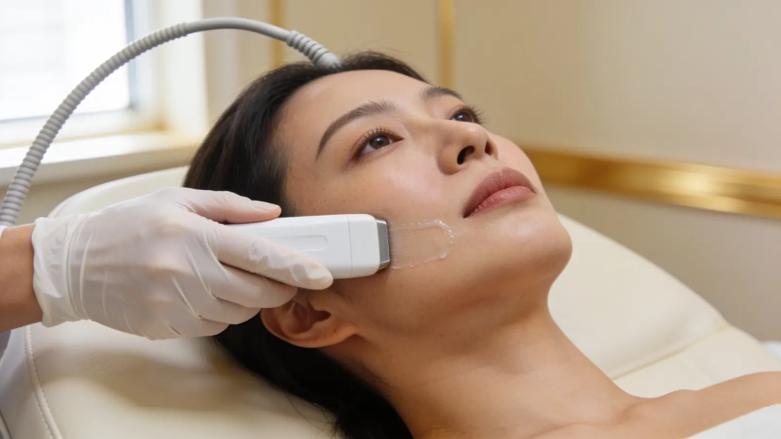
Cheek filler is often placed in more than one layer, from the bone to just under the skin, and the deposit you can feel may not be the one causing the fullness. Ultrasound shows where the filler sits, how deep it is and how far it has spread, and what it is likely made of. It also shows the blood vessels nearby, which guides where a needle goes.
Illustrative image — not a patient.
Different fillers tend to look different on the scan. In a study of 60 people with previous fillers, 48 showed a well-defined pattern and 12 a mixed one, mainly from different procedures at different times. Liquid silicone looked like fine-grain ‘snowfall’, calcium hydroxylapatite and PMMA like coarse-grain snowfall, and polyacrylamide and polyalkylimide gels, like hyaluronic acid soon after injection, appeared as rounded, cyst-like pockets (Urdiales-Gálvez 2021). An international consensus of 15 experts from seven specialties calls ultrasound mandatory for managing filler complications, and recommends identifying fillers already in place before any further injection (Velthuis 2025).
The scan shows what a lump is likely made of; it does not prove it. Where something does not look like filler, such as a cyst or a lymph node, further tests are arranged. Sometimes the scan shows that what looked like filler is fluid, fat or loose skin, and nothing needs dissolving at all. Dr Sin Yong performs the scan himself, and the plan follows what it shows. More on scanning is in ultrasound-guided filler correction.
What Dr Sin Yong does instead of cutting cheek filler out
Maps. He scans both cheeks himself with ultrasound: the likely material, its depth, how far it has spread and the vessels nearby.
Treats inflammation or infection first. A red, hot, tender, swollen or draining lump is treated medically before any dissolving or laser.
Dissolves hyaluronic acid. He uses hyaluronidase, an enzyme that acts on hyaluronic acid only. Your allergy history is checked, including any reaction to bee or wasp stings and any earlier hyaluronidase. Before planned dissolving, Dr Sin Yong always does a skin test: a small amount of hyaluronidase is injected just under the skin to check for a reaction. A small first dose goes into the mapped excess, with a review at about 48 hours, because dissolving more than the excess can leave the cheek flat or hollow. Different hyaluronic acid fillers respond differently to the enzyme, so doses are staged. In an emergency such as a blocked blood vessel, dissolving is never delayed for a skin test.
Treats what cannot be dissolved, without an incision. He treats Sculptra, Radiesse, Ellansé, Juvelook, silicone and other non-absorbable fillers, layered products and excess fat graft with the Sculpting mode of Time Freeze Laser LCLR®, his own protocol on a long-pulsed 755 nm and 1064 nm laser platform, directed at the mapped material. Like any laser, it can cause blistering or a change in skin colour, so settings are chosen for your skin. On the upper cheek, close to the eye, settings are kept conservative and the eyes are protected.
Reviews. The cheeks are judged once they have settled, not on the day, and any further visit is planned from what is seen.
He does not add filler to hide filler. If you want filler again, it waits at least two weeks after hyaluronidase and comes only after the excess has been dealt with; sometimes the answer is less filler, placed differently, and sometimes none. Where loose skin is part of the picture, lifting is considered separately, after any excess filler has been addressed. Dissolving is set out step by step in cheek filler dissolving, the skin test in hyaluronidase allergy and the skin test, and the laser in the Sculpting mode of Time Freeze Laser LCLR®.
Surgery to remove cheek filler: what it involves
Surgery to remove filler from the cheek means cutting it out, through the skin of the face or from inside the mouth, or drawing it out through a small opening. Each route has its own trade-offs.
Swipe sideways to see the whole table.
Approach
What it involves
Incision
What to weigh up
Excision through the skin
Cutting out the lump together with the tissue around it
Yes, on the face
A visible scar; a dent where tissue is removed; nerve injury; product left behind; cannot be undone
Excision from inside the mouth
Reaching the cheek through the lining of the mouth
Yes, inside the mouth
No scar on the skin, but tissue is still cut, with the same risks of a dent, nerve injury and product left behind
Removal through facelift incisions
Reaching the filler through incisions around the ear, as part of a facelift
Yes, around the ear
Scars around the ear; nerve injury; a facelift lifts skin and deeper tissue, but on its own it does not take filler out
Needle aspiration or squeezing out
Drawing soft gel out through a needle or a small opening
A needle puncture or a small opening
Possible only while the gel is soft and gathered, not once it is woven into scar tissue
Hyaluronidase
Enzyme injected into mapped hyaluronic acid
None; fine needle
Acts on hyaluronic acid only; skin test first; too much can leave a hollow
Sculpting mode of Time Freeze Laser LCLR®
Laser directed at mapped material that cannot be dissolved
None
Can cause blistering or a change in skin colour, so settings are chosen for your skin
One published series shows why taking filler out of the face is harder than it sounds. In one series of 31 patients operated on for contour problems after facial filler, by excision through facelift incisions, needle aspiration or excision inside the mouth, scar tissue and the reaction around the filler made the dissection difficult; 1 had a facial nerve injury, which improved after four months, 2 had collections of blood under the skin and 1 a collection of fluid (Ghareeb 2022).
Surgery has a place. The same authors found the facelift approach more demanding but more satisfying for major deformities after repeated injections (Ghareeb 2022). An abscess may need draining, and a lump that does not look like filler on the scan may need a tissue sample. The point is the order: the decision to operate should follow an ultrasound assessment, not come before it. An operation can still be done later if it is ever needed; it cannot be undone. The operations and their published complications are covered in filler removal surgery.
Warning signs after cheek filler: when to act at once
A blocked blood vessel is a rare but recognised risk of cheek filler. Arteries in the cheek, especially towards the side of the nose and below the eye, connect with the vessels that supply the eye, which is why a change in vision after cheek filler is an emergency. A blocked vessel usually shows during or soon after the injection, sometimes later: act whenever it appears.
Urgent: a change in vision or eye pain after filler: call 995 now. Skin that turns pale, dusky or mottled, or pain that is getting worse, needs urgent assessment by the doctor who injected you, or at A&E, now. See the warning signs of a blocked blood vessel after filler.
Skin that is dying after a blocked blood vessel (necrosis) is an emergency. It needs urgent, more intensive care than a lump does, starting now.
A lump or swelling that is red, hot, tender or draining may be inflammation or infection. It is treated medically first, before any dissolving, any laser and any talk of surgery. Fever, or feeling unwell with a swollen lump, needs review the same day.
Difficulty breathing, a tight throat, swelling of the tongue or throat, or swelling that spreads quickly across the face, after any injection, including hyaluronidase, can be a serious allergic reaction: call 995 now. Itch, hives or swelling that spreads beyond the treated area: contact the clinic that injected you the same day.
Do not press hard on a cheek lump, massage it firmly or try to break it up yourself.
Tried other treatments and almost given up?
Many patients who come to see Dr Sin Yong about cheek filler that has gone wrong tell him they have tried almost everything else and had given up hope. Having tried other treatments does not mean nothing more can be done: what is done next depends on what the scan shows.
That history is useful. Knowing what was injected, when, and what has already been tried often explains why a problem has stayed: hyaluronidase acts only on hyaluronic acid and only where it reaches, some fillers are not hyaluronic acid at all, and swelling or fluid can look like filler. Ultrasound helps show which of these applies, and the plan starts from there. For material that cannot be dissolved, Dr Sin Yong uses the Sculpting mode of Time Freeze Laser LCLR® for nodules large and small, without an incision. How each person responds varies.
If you have been told nothing more can be done, it is still worth having the area scanned: ultrasound helps show what is there, and the plan is made from that. Bring any records of past treatments, product names and dates if you have them. More on this is in when filler dissolving has not worked.
How Dr Sin Yong treats cheek filler gone wrong
Dr Sin Yong developed the Time Freeze Laser LCLR® protocol himself and performs every scan, every dissolving injection and every Sculpting mode treatment himself on Orchard Road. He has been invited to share his clinical expertise in Seoul, Dubai, Bangkok and São Paulo, including as a Global Key Opinion Leader.
He examines the cheeks at rest and when you smile, from the front and the side, because a problem in the cheek changes how the under-eye and the smile lines look. He explains the risks and limits of each option at the assessment, including waiting where that is reasonable and surgery where it is needed. You do not need to have been treated here, and having no records of what was injected is fine; filler placed overseas is assessed in the same way, as set out in correction of filler done overseas. How cheek filler itself is planned is covered in cheek fillers.
Your next step
Not for emergencies. A change in vision or eye pain after filler: call 995 now. Pale, dusky or mottled skin, or pain that is getting worse: go now to the doctor who injected you, or to A&E. Do not message or book for these signs.
1. MessageWhatsApp +65 8023 7170 with what is bothering you. A reply comes from the clinic, not a bot.
2. AssessmentDr Sin Yong examines you himself on Orchard Road, with ultrasound where earlier filler is involved.
3. A plan in writingWhat he would do, what he would not, the fee factors, and the option of no treatment.
Every treatment is performed by Dr Sin Yong personally. Singapore Medical Council registered · invited to share his clinical expertise internationally for CLASSYS, DEKA, Fotona, Hironic and Ilooda.
Usually not. Get assessed first. Hyaluronic acid is dissolved, and Dr Sin Yong treats Sculptra, Radiesse, Ellansé, silicone and other material that does not dissolve with the Sculpting mode of Time Freeze Laser LCLR®, without an incision. Surgery leaves a scar, can leave a dent and can injure the nerves that move your smile or give the cheek feeling. In his practice, almost every hard lump is treated without surgery. If yours is one of the few that needs surgery, you are told plainly, and why.
Many of the patients Dr Sin Yong sees have tried other treatments first. A treatment that did not help can still leave options open: hyaluronidase may not have reached the filler, the filler may not be hyaluronic acid, or what looks like filler may be swelling or fluid. Ultrasound helps show which applies, and the plan follows what it finds.
Usually one or more of these: cheeks that look heavy, round or too wide and rise as a block when you smile; puffiness on the upper cheek below the eyes; fullness that has slid into the smile lines or up towards the lower eyelid; lumps you can see or feel; or one cheek fuller than the other once the swelling has gone. Ultrasound shows which of these is present and what the filler is likely made of, and the plan follows from that.
Usually because the cheeks are carrying more filler than they can hold. Hyaluronic acid lasts longer than many people expect, so top-ups are often added to filler that is still there: in a review of 1,410 patients, 3D imaging found 50–86% still present at 24 months (de Castro Costa 2025). Some filler may also expand after it is placed (Issa 2026). Puffiness below the eyes can be malar oedema. The scan shows which, and the hyaluronic acid excess is dissolved in stages.
It can. Filler placed high on the cheek, or filler that has moved up towards the lower eyelid, can hold fluid in an area that drains poorly, called malar oedema. Expert recommendations list a high volume of filler and placement superficial to the malar septum among the risks (Baranska-Rybak 2026). In 61 patients with long-lasting lower-eyelid swelling after hyaluronic acid filler to the lower eyelids or cheeks, one hyaluronidase treatment was effective in 92% (Skippen 2020). The scan tells filler from fluid first.
First, find out where it is now. Cheek filler can slide lower or forwards, spread into the smile lines or creep up towards the lower eyelid, sometimes long after the injection. Ultrasound shows where it sits and what it is likely made of. Hyaluronic acid that has moved is dissolved where it lies, after a skin test, and filler still doing useful work can stay. Material that cannot be dissolved is treated with the Sculpting mode of Time Freeze Laser LCLR®, without an incision. Do not massage it.
They are cheeks carrying more filler than they can hold naturally. They sit too high and too round, rise as a block when you smile, push the lower eyelids up and make the face look wider. A review of facial overfilled syndrome describes ‘chipmunk’ cheeks, attributes the syndrome to excessive filler and poor technique, and notes that it is reported mainly in people of East Asian descent (Lim 2024). Hyaluronic acid excess is mapped and dissolved in stages, aiming at the excess rather than every trace.
It depends on the filler. Hyaluronic acid lumps are usually product gathered in one spot or too close to the surface. Collagen stimulators are often placed in the cheeks: in 130 people treated with Sculptra, nodules were the commonest side effect (8.5%), most often in the hands and cheeks (Palm 2010). Silicone and other non-absorbable fillers from older treatments can form lumps years later. A lump that is red, hot or tender is treated medically first, and ultrasound shows what a lump is likely made of.
Early swelling and tenderness over the cheekbones are expected, sometimes with bruising, and normal swelling eases steadily. It is a problem when it grows instead of easing, when puffiness stays below the eyes or on the upper cheek, when the face looks rounder at rest than before, or when swelling settles and then comes back. In one case series, malar oedema after hyaluronic acid filler began anywhere from immediately to three years after treatment (Schelke, J Cosmet Dermatol 2023). Swelling that will not settle is scanned; swelling with pale, dusky or mottled skin, worsening pain or any change in vision is an emergency.
No. Poly-L-lactic acid (Sculptra), calcium hydroxylapatite (Radiesse) and polycaprolactone (Ellansé) do not respond to hyaluronidase, which acts on hyaluronic acid only. That does not mean they have to be cut out. Dr Sin Yong maps them with ultrasound and treats them with the Sculpting mode of Time Freeze Laser LCLR®, for nodules large and small, without an incision. Where hyaluronic acid has been layered over them, that part is dissolved first.
No. Filler added around a lump or a heavy cheek hides the problem instead of dealing with it, adds weight to a face that is already carrying too much, and layers new product over old, which makes the next scan and the next treatment harder. In one MRI study of 14 people, hyaluronic acid filler measured about 2.8 times the volume injected (Issa 2026). Deal with the excess first. Whether anything is added back is decided once the cheeks have settled, and sometimes the answer is nothing.
A scar, on the face or inside the mouth; a dent where tissue is removed; injury to the branches of the facial nerve that move the smile, or to the nerve that gives feeling to the cheek and upper lip; and filler left behind. In 31 patients operated on after facial filler, scar tissue made the dissection difficult, 1 had a facial nerve injury that improved after four months, and 2 had collections of blood (Ghareeb 2022). Surgery cannot be undone, so get assessed first.
Not reliably. Hyaluronic acid often lasts far longer than people expect, so overfill and filler that has moved tend to stay: ultrasound has shown hyaluronic acid 12 to 36 months after injection (de Castro Costa 2025). Collagen stimulators cannot be dissolved, and the collagen they provoke can last for years; silicone and other non-absorbable fillers stay for life. Both can still be treated without an incision. A small change that does not bother you can reasonably be watched, and the scan helps decide whether that is sensible.
A change in vision or eye pain after filler is an emergency: call 995 or go to the nearest emergency department now. Skin on the cheek or beside the nose that turns pale, dusky or mottled, or pain that is getting worse, needs urgent assessment by the doctor who injected you, or at A&E. A lump that is red, hot, tender or draining, especially with a fever, needs medical review the same day. Skin that is dying after a blocked vessel needs urgent, more intensive care, starting the same day.
References
Exploring facial overfilled syndrome from the perspective of anatomy and the mismatched delivery of fillers. Journal of Cosmetic Dermatology, 2024. source
Avoiding malar edema during midface/cheek augmentation with dermal fillers. Journal of Clinical and Aesthetic Dermatology, 2011. source
Extratemporal Facial Nerve: Surgical Landmarks, Risk Zones, and a Practical Framework for Safe Dissection. Laryngoscope Investigative Otolaryngology, 2026. source
Complications following Permanent Filler Injection: A Prospective Cohort Study and Protocol of Management. Plastic and Reconstructive Surgery Global Open, 2022. source
Cosmetic use of poly-l-lactic acid: a retrospective study of 130 patients. Dermatologic Surgery, 2010. source
Longevity and Volume Expansion of Hyaluronic Acid Dermal Fillers: A Cross-sectional 3-dimensional Magnetic Resonance Imaging Study. Plastic and Reconstructive Surgery Global Open, 2026. source
The use of ultrasound imaging in aesthetic injectables: A modified Delphi consensus. Journal of Plastic, Reconstructive & Aesthetic Surgery, 2025. source
Facial Overfilled Syndrome: A Narrative Clinical Review. Clinical, Cosmetic and Investigational Dermatology, 2026. source
Clinical Durability of Hyaluronic Acid-Based Dermal Fillers for Facial Application: A Systematic Review. Aesthetic Plastic Surgery, 2025. source
Malar Edema after Hyaluronic Acid Dermal Fillers: Expert Recommendations on Etiology, Prevention, and Management. Dermatology and Therapy, 2026. source
Periorbital venous stasis may be involved with filler induced malar edema: a duplex ultrasound-imaging-based case series. Journal of Cosmetic Dermatology, 2023. source
Rehabilitation of the Dysmorphic Lower Eyelid From Hyaluronic Acid Filler: What to Do After a Good Periocular Treatment Goes Bad. Aesthetic Surgery Journal, 2020. source
Calcium hydroxylapatite filler: an overview of safety and tolerability. Journal of Drugs in Dermatology, 2013. source
Ultrasound patterns of different dermal filler materials used in aesthetics. Journal of Cosmetic Dermatology, 2021. source
Complicated Facial Fillers: Management Algorithm. Plastic and Reconstructive Surgery Global Open, 2022. source
About this page: written and medically reviewed by Dr Sin Yong, MBBS (NUS), MRCS (Edinburgh) · Last reviewed 26 October 2026 · Illustrative images only, not patients · Editorial policy
Not for emergencies. A change in vision or eye pain after filler: call 995 now. Pale, dusky or mottled skin, or pain that is getting worse: go now to the doctor who injected you, or to A&E. Do not message or book for these signs.