Published 26 October 2026 · Reviewed by Dr Sin Yong
Many patients who see Dr Sin Yong about filler problems tell him they have tried almost everything else and had given up hope. When filler seems to remain after hyaluronidase, the reasons can be looked for: the filler may not be hyaluronic acid, the enzyme may not have reached it, or the fullness may be swelling, fluid or your own tissue.
Short answerWhen filler remains after hyaluronidase, the usual explanations are that the material is not hyaluronic acid (Sculptra, Ellansé, Radiesse and the poly-D,L-lactic acid in Juvelook do not dissolve), that the enzyme did not reach a deep or dense deposit, that a staged dose has not yet been reviewed, or that the fullness is swelling, fluid or tissue. Ultrasound helps show which applies (Wortsman 2015). Hyaluronic acid that remains can be targeted again under ultrasound; for nodules large and small that cannot be dissolved, Dr Sin Yong uses the Sculpting mode of Time Freeze Laser LCLR®.
Many patients who come to see Dr Sin Yong about filler problems tell him they have tried almost everything else and had given up hope.
Having tried other treatments does not, on its own, rule out further options. What can be done depends on what the scan shows: what the material is, where it sits and how much of it is left. That is why a second look starts with ultrasound rather than with another dose of enzyme.
60-second filler second-look check
Answer three questions and send them straight to the clinic on WhatsApp. Dr Sin Yong will let you know whether an ultrasound assessment is worthwhile. No obligation.
1 · When was the filler done?
2 · What bothers you most?
3 · Have you already tried other treatments for this?
Your answers open in your own WhatsApp app; nothing is stored on this page.
Why filler can remain after hyaluronidase
Hyaluronidase is an enzyme that breaks down hyaluronic acid. It does nothing to other fillers, and it only acts where it reaches. Published reviews also note that hyaluronic acid products differ in how readily the enzyme breaks them down, which is one reason doses are staged and reviewed (Landau 2015; Kroumpouzos and Treacy 2024). The table sets out the common reasons filler seems to stay.
Swipe sideways to see the whole table.
Reason
What you may notice
What ultrasound can help show
What is considered
The filler is not hyaluronic acid
Little or no change; a firm lump stays
Material that does not look like hyaluronic acid; collagen stimulators have their own appearances (Wortsman 2015)
Not dissolved; for nodules large and small, the Sculpting mode of Time Freeze Laser LCLR®
The enzyme did not reach the deposit
Part of the area softened, part did not
A deposit deeper than, or to one side of, where the enzyme went
Hyaluronidase placed into the deposit under ultrasound (Quezada 2016)
Dense or well-defined hyaluronic acid
A firm lump that softens only partly
A compact deposit with clear edges
Targeted doses into the deposit, staged and reviewed
The review is not yet due
Swelling makes the result hard to judge in the first days
Swelling around a smaller deposit
Review at about 48 hours before more is added (King 2018)
Swelling, fluid or tissue rather than filler
Puffiness that changes through the day, often under the eyes or over the cheeks
Fluid or tissue without a filler deposit
The cause of the swelling is assessed; more enzyme is not the answer
Layered or mixed products
Lumps in several areas after years of treatment
Different materials at different depths
Hyaluronic acid targeted; the rest managed as collagen-stimulator material
Filler that has been in place for years can also change. Hyaluronic acid breaks down slowly and unevenly, and other materials are cleared by the body through different, slower processes (Wollina 2024). A lump that appears months or years later is a symptom rather than a diagnosis, and a full history and assessment come before any treatment (Convery 2021).
“A biostimulator cannot be dissolved. That single fact should shape every decision about using one.”
Dr Sin YongOn why some fillers do not dissolve
What a second look with ultrasound shows
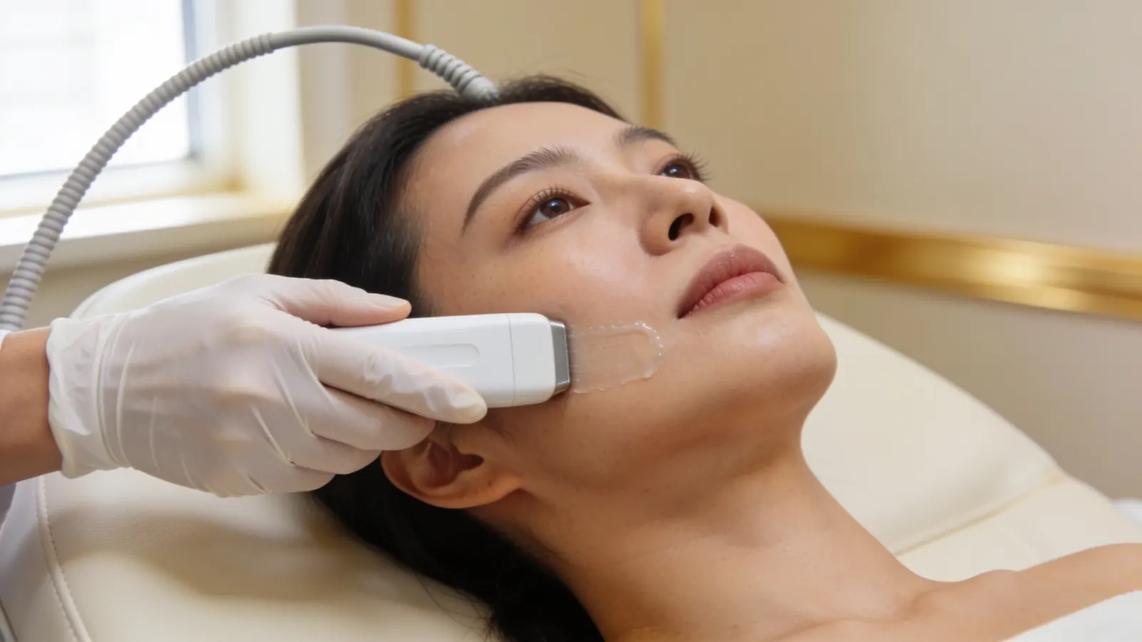
High-frequency ultrasound shows the layers of the face and what sits between them. Hyaluronic acid, calcium hydroxylapatite and polycaprolactone tend to look different on the scan, and the scan shows how deep a deposit sits and whether blood vessels run close to it (Wortsman 2015; Schelke 2018). Dr Sin Yong performs the scan and any injection himself.
Illustrative image — not a patient.
Seeing the deposit changes how the enzyme is used. In a study of blocked-vessel emergencies, placing hyaluronidase into the filler seen on ultrasound used a mean of 122.5 units, against 1,519.4 units when the whole region was flooded (Schelke, Aesthet Surg J 2023). That study was about emergencies, but the principle of aiming at what can be seen applies to planned dissolving too.
When hyaluronic acid remains: targeting it again
If the scan shows a deposit that looks like hyaluronic acid, it can be dissolved again, this time into the deposit itself. A small dose is placed, the area is reviewed at about 48 hours, and more is added only where filler remains (King 2018). Allergy history is checked again, including any reaction to earlier doses and any bee or wasp sting allergy, with a skin test where there is concern.
Dissolving more than was in excess can leave an area looking hollower than expected, which is why the dose is staged. Refilling, if it is wanted at all, waits at least two weeks and until the swelling has settled (Murray 2021). The steps are set out in filler dissolving in Singapore, with what to expect afterwards in after filler dissolving.
When the filler cannot be dissolved
Sculptra (poly-L-lactic acid), Ellansé (polycaprolactone), Radiesse (calcium hydroxylapatite) and Juvelook (poly-D,L-lactic acid) are not broken down by hyaluronidase (Lin and Christen 2020; Wollina 2024); in Juvelook only the hyaluronic acid carrier is. If one of these was treated as hyaluronic acid, the enzyme had nothing to act on. Nodules after collagen-stimulating fillers are described in a published case series, and they behave differently from hyaluronic acid lumps (Ianhez 2024).
Not every fullness after filler is filler. Swelling over the cheeks or under the eyes can come from fluid that the tissue holds. Published imaging work suggests that filler pressing on the veins and lymphatic drainage around the eye can play a part; in that case series the swelling improved after the filler was removed under ultrasound guidance (Schelke, J Cosmet Dermatol 2023). Eye bags, loose skin, fat and the natural shape of the face can all look like leftover filler, particularly in photographs taken in overhead light.
Dissolving cannot change eye bags, loose skin, fat or facial shape, so the examination and scan come first. Where the cause is laxity or fat rather than filler, the options are different, and they are discussed at the same visit. See under-eye filler dissolving and pillow face.
What to bring to a second assessment
Product names, sticker cards or invoices from earlier treatments, if you have them.
Approximate dates of each filler treatment and each dissolving visit.
Which areas were treated, and with what, as far as you know.
Any reaction to earlier hyaluronidase, and any allergies.
Photographs from before any filler, which help show your own facial shape.
Urgent: a change in vision or eye pain after filler means calling 995 or going to the nearest emergency department now. Skin that turns pale, dusky or mottled, or pain that is getting worse, needs urgent assessment by the doctor who injected you, or at A&E. See the warning signs of a blocked blood vessel after filler.
A lump that becomes red, hot, painful or quickly larger may be inflamed or infected and needs a prompt medical review rather than a planned dissolving visit.
How Dr Sin Yong approaches a second look
Dr Sin Yong sees every patient himself on Orchard Road, scans before anything is planned and performs any injection or laser treatment himself. He does not add more product to hide a lump, and he does not repeat a treatment that has already failed without first finding out why it did.
He developed the Time Freeze Laser LCLR® protocol and has been invited to share his clinical expertise internationally, including as a Global Key Opinion Leader. Whatever the history, the plan follows what the scan shows, and how each person responds varies.
Your next step
1. MessageWhatsApp +65 8023 7170 with what is bothering you. A reply comes from the clinic, not a bot.
2. AssessmentDr Sin Yong examines you himself on Orchard Road, with ultrasound where earlier filler is involved.
3. A plan in writingWhat he would do, what he would not, the fee factors, and the option of no treatment.
Every treatment is performed by Dr Sin Yong personally. Singapore Medical Council registered · invited to share his clinical expertise internationally for CLASSYS, DEKA, Fotona, Hironic and Ilooda.
The common reasons are that the filler was not hyaluronic acid, that the enzyme did not reach a deep or dense deposit, that a staged dose had not yet been reviewed, or that the fullness was swelling, fluid or your own tissue rather than filler. Ultrasound helps show which of these applies before anything more is injected.
Many of the patients Dr Sin Yong sees have tried other treatments first. A treatment that did not help can still leave options open: hyaluronidase may not have reached the filler, the filler may not be hyaluronic acid, or what looks like filler may be swelling or fluid. Ultrasound helps show which applies, and the plan follows what it finds. What can be done varies from person to person.
Hyaluronidase breaks down hyaluronic acid, but only where it reaches and only if the filler is hyaluronic acid. Products also differ in how readily the enzyme acts on them, so a single dose can leave a deposit behind. That is a reason to look again with ultrasound, not necessarily a reason to stop.
The enzyme starts acting soon after it is placed, but swelling in the first days hides the result. The area is usually judged at a review about 48 hours later, and a firm view is easier once the swelling has fully settled. If a lump is still there after that, have it scanned.
Yes, if the scan shows a deposit that looks like hyaluronic acid. The second round is aimed at the deposit itself, with a small dose, a review at about 48 hours and more only where filler remains. Allergy history is checked again, including any reaction to earlier doses.
These are collagen stimulators and hyaluronidase does not dissolve them. They are mapped with ultrasound, and for nodules large and small Dr Sin Yong uses the Sculpting mode of Time Freeze Laser LCLR®, without an incision. How each person responds varies.
Sometimes. Swelling from the injections themselves settles over days, and some puffiness under the eyes or over the cheeks comes from fluid rather than filler. Ultrasound helps tell fluid and tissue apart from a filler deposit, which matters because more enzyme will not help swelling that is not caused by filler.
Staged doses are a normal part of dissolving. Each dose carries risks, including bruising, swelling and, uncommonly, an allergic reaction. Earlier hyaluronidase injections are one of the main risk factors for allergy, and many reported reactions happened at a second injection (Guliyeva 2024), so any reaction to an earlier dose is reported before another is given. Dissolving more than is in excess can leave an area looking hollow, which is why each step is reviewed.
It can still be worth having the area scanned: ultrasound helps show what is still there and where it sits, which is what any decision, including no treatment, depends on. Many patients who see Dr Sin Yong have tried other treatments first, and the answer varies from person to person.
They help but are not required. Product names, dates and the areas treated make the scan easier to interpret. Photographs from before any filler help show your own facial shape.
Yes. Filler placed at another clinic in Singapore or overseas is assessed in the same way: your history, an examination and an ultrasound scan first, then a plan based on what is found. Product names and dates help if you have them, but the scan does not depend on them, and you do not need a referral to be seen.
References
Identification and Complications of Cosmetic Fillers: Sonography First. Journal of Ultrasound in Medicine, 2015. source
Ultrasound to improve the safety of hyaluronic acid filler treatments. Journal of Cosmetic Dermatology, 2018. source
Ultrasound-guided hyaluronidase injection in cosmetic complications. Journal of the European Academy of Dermatology and Venereology, 2016. source
Ultrasound-Guided Targeted vs Regional Flooding: A Comparative Study for Improving the Clinical Outcome in Soft Tissue Filler Vascular Adverse Event Management. Aesthetic Surgery Journal, 2023. source
The Use of Hyaluronidase in Aesthetic Practice (v2.4). Journal of Clinical and Aesthetic Dermatology, 2018. source
Guideline for the Safe Use of Hyaluronidase in Aesthetic Medicine, Including Modified High-dose Protocol. Journal of Clinical and Aesthetic Dermatology, 2021. source
Hyaluronidase Caveats in Treating Filler Complications. Dermatologic Surgery, 2015. source
Hyaluronidase for Dermal Filler Complications: Review of Applications and Dosage Recommendations. JMIR Dermatology, 2024. source
Periorbital venous stasis may be involved with filler induced malar edema: a duplex ultrasound-imaging-based case series. Journal of Cosmetic Dermatology, 2023. source
Spontaneous and induced degradation of dermal fillers: A review. Journal of Cutaneous and Aesthetic Surgery, 2024. source
Delayed-onset Nodules (DONs) and Considering their Treatment following use of Hyaluronic Acid (HA) Fillers. Journal of Clinical and Aesthetic Dermatology, 2021. source
Polycaprolactone-based dermal filler complications: A retrospective study of 1111 treatments. Journal of Cosmetic Dermatology, 2020. source
Complications of collagen biostimulators in Brazil: Description of products, treatments, and evolution of 55 cases. Journal of Cosmetic Dermatology, 2024. source
Allergic Complications of Hyaluronidase Injection: Risk Factors, Treatment Strategies, and Recommendations for Management. Aesthetic Plastic Surgery, 2024. source
About this page: written and medically reviewed by Dr Sin Yong, MBBS (NUS), MRCS (Edinburgh) · Last reviewed 26 October 2026 · Illustrative images only, not patients · Editorial policy