Published 13 November 2026 · Reviewed by Dr Sin Yong
A fibrotic filler lump is a firm lump made partly or wholly of scar tissue that the body has laid down around injected filler. It feels harder than the filler did, it does not soften with massage, and dissolving injections alone do not clear it, because they act on hyaluronic acid and not on scar tissue.
Short answerA fibrotic filler lump is scar tissue (fibrosis) the body has formed around or in place of filler. It tends to follow repeated treatment in one area, layered products or earlier inflammation. A finger cannot tell fibrosis from a lump of product or a granuloma; ultrasound can help. Hyaluronidase dissolves hyaluronic acid only, so any product inside the lump and the fibrous tissue are assessed separately. Dr Sin Yong scans each lump himself on Orchard Road, Singapore, before advising treatment.
Fibrosis is the body’s repair response: it lays down collagen-rich scar tissue. Around injected filler, that tissue can form a firm shell, run through the product, or remain after the product itself has gone. The result is a lump that is harder than filler and does not behave like it.
It is one of several things a hard lump can be. Others are filler collected in one place, a filler granuloma, and an inflamed or infected nodule. They can feel alike under a finger and can exist together in the same lump. The wider picture is on filler lumps and nodules.
Why does fibrosis form after filler?
Fibrosis follows repeated or prolonged irritation of tissue. With filler, the usual settings are repeated treatment in the same area over years, different products layered on top of each other, a past episode of swelling or infection, and the body walling off material it cannot break down.
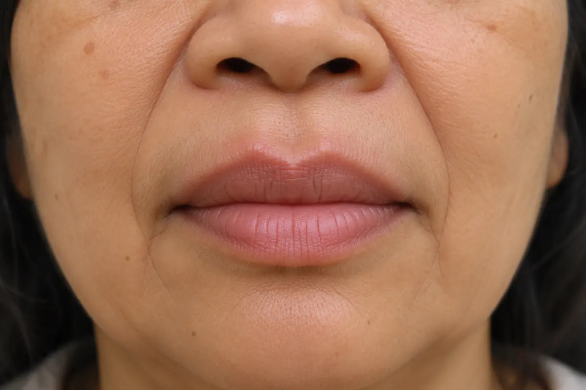
Fullness above the lip border can be filler that has moved. Illustrative image, not a patient.
In a small ultrasound study of lumps inside the lips after filler, the two cases of marked fibrosis were attributed to repeated filler procedures, including fillers other than hyaluronic acid and earlier attempts to remove filler or treat complications (Skrzypek and Mlosek 2020).
Collagen-stimulating products work by prompting the body to make collagen, so a nodule from those products is a related but separate problem. Those are covered on Sculptra lumps, Ellansé lumps and Juvelook lumps.
Fibrotic lump, filler lump, granuloma or inflamed nodule?
The four are managed differently, so telling them apart is the first step.
Swipe sideways to see the whole table.
What it is
Typical timing
Does hyaluronidase clear it?
Lump of filler
The product itself, gathered in one place or placed too near the surface
Usually soon after treatment
Yes, where the product is hyaluronic acid
Fibrotic lump
Scar tissue around or in place of the filler
Usually late; often after repeated treatment
No; only any hyaluronic acid still inside it
Granuloma
An immune reaction that walls the filler off, often with scar tissue
Usually months to years later
Only where the filler is hyaluronic acid, and often not on its own
Inflamed or infected nodule
A red, hot, tender or draining lump
Any time
Not the first step; it is treated medically first
These are general patterns, not rules, and one lump can contain more than one of them.
How is a fibrotic lump identified?
By the history, the examination and a scan. In 15 women with lumps where hyaluronic acid filler had been injected, high-frequency ultrasound showed filler deposits in 9, granulomas in 3, fibrosis in 2 and a deposit with inflammation in 1, and the authors reported that each had a typical appearance on the scan (Mlosek 2020).
A scan also shows how deep the lump sits, how far it extends and which vessels run beside it. Where the picture is unclear, or the lump does not look like filler at all, further tests including a tissue sample are arranged. How Dr Sin Yong uses the scan is described under ultrasound-guided filler dissolving.
Why does dissolving not clear a fibrotic lump?
Hyaluronidase is an enzyme that breaks down hyaluronic acid. It does not act on scar tissue, and it does not act on fillers made of other materials. If a lump is still there after dissolving, the possibilities include hyaluronic acid that the enzyme did not reach, a filler that is not hyaluronic acid, a granuloma, or fibrous tissue.
An international consensus on delayed reactions to hyaluronic acid filler also notes that hyaluronidase can fail on its own, particularly with firmer, more tightly cross-linked gels (Philipp-Dormston 2020). Repeating it without knowing what the lump is made of is not a plan. The causes are set out on hyaluronidase not working.
What does published guidance describe for lumps that persist?
The same consensus groups late lumps into inflamed and non-inflamed, and describes these steps (Philipp-Dormston 2020):
Non-inflamed lumps: observation is acceptable; hyaluronidase where hyaluronic acid is present.
Inflamed lumps: antibiotics as the main approach, with anti-inflammatory medicine or corticosteroid in selected cases, and drainage of an abscess.
Lumps that do not respond: corticosteroid with 5-fluorouracil injected into the lump itself, not into the skin over it, because steroid in the skin can thin it.
Surgical removal: a last resort.
That guidance was written about hyaluronic acid filler and does not address fibrosis as a separate entity. It is given here as background, not as a treatment plan for any individual.
When is a hard lump urgent?
Urgent: a change in vision or eye pain after an injection means calling 995 now. Skin that turns white, pale, dusky or mottled, or pain that is getting worse, needs the doctor who injected you, or A&E, now.
A lump that becomes red, hot, swollen, tender or starts to drain needs a doctor the same day, or A&E if no doctor can see you. Infection and inflammation are treated medically before anything else is done to the lump.
How Dr Sin Yong assesses a fibrotic filler lump
History. What was injected, where and when, if known; what has been tried, including dissolving; what you were offered. Having no records is fine.
Ultrasound mapping. He scans the lump himself: what is likely to be product, what is likely to be fibrous tissue, how deep it sits and which vessels are nearby.
Inflammation first. A red, hot, tender or draining lump is treated medically before anything else.
Product inside the lump. Hyaluronic acid is dissolved under ultrasound guidance after a skin test. Filler that cannot be dissolved is treated with the Sculpting mode of Time Freeze Laser LCLR®, directed at the mapped lump, without an incision.
The fibrous tissue. What remains is reassessed on the scan. He explains the options for it and their limits, including a surgical opinion where that is the appropriate route, and leaving it alone where it is small and stable.
Review. Each lump is reassessed, and any further visit is planned from what is seen.
He does not add more filler to hide a lump. Like any laser, the Sculpting mode can cause blistering or a change in skin colour, so settings are chosen for your skin.
Only offered surgery for a hard lump?
Cutting a lump out leaves a scar and can leave a dent, and removal is often incomplete where the tissue has no clear border. What published series report, both where surgery fell short and where it did well, is set out on filler granuloma. An assessment with a scan first shows what the lump is made of, which is what any decision about surgery should rest on.
Your next step
Not for emergencies. A change in vision or eye pain after an injection: call 995 now. Pale, dusky or mottled skin, or pain that is getting worse: go now to the doctor who injected you, or to A&E. Do not message or book for these signs.
1. MessageWhatsApp the clinic with what is bothering you, and the product and date if you know them. A reply comes from the clinic, not a bot.
2. AssessmentDr Sin Yong examines you himself on Orchard Road, with an ultrasound scan of the area.
3. A plan in writingWhat he would do, what he would not, the fee factors, and the option of no treatment.
Every treatment is performed by Dr Sin Yong personally. Singapore Medical Council registered · invited to share his clinical expertise internationally for CLASSYS, DEKA, Fotona, Hironic and Ilooda.
A hard lump is examined and scanned with ultrasound before any treatment is advised. How consultations work.
A fibrotic filler lump is a firm lump made partly or wholly of scar tissue (fibrosis) that the body has formed around or in place of injected filler. It is harder than the filler was and does not soften with massage.
Not by hyaluronidase alone. Hyaluronidase breaks down hyaluronic acid and does not act on scar tissue. Any hyaluronic acid still inside the lump can be dissolved; the fibrous tissue is assessed separately.
A finger cannot tell reliably. History, examination and high-frequency ultrasound help to tell filler deposits, granulomas, fibrosis and inflammation apart. Where the scan is unclear, a tissue sample may be arranged.
Possible reasons are hyaluronic acid the enzyme did not reach, a filler that is not hyaluronic acid, a granuloma, or fibrous tissue. Repeating hyaluronidase without knowing which applies is not advised; a scan comes first.
A stable firm lump that does not hurt is not an emergency. A lump that turns red, hot, swollen, tender or starts to drain needs a doctor the same day. Skin colour change or a change in vision after an injection is an emergency.
Not always. It depends on what the scan shows the lump is made of, its size and depth, and whether it is changing. Surgery leaves a scar and removal can be incomplete, so an assessment with ultrasound comes before that decision.
References
Mlosek RK, Migda B, Skrzypek E, Słoboda K, Migda M. The use of high-frequency ultrasonography for the diagnosis of palpable nodules after the administration of dermal fillers. Journal of Ultrasonography, 2020. source
Skrzypek E, Mlosek RK. High frequency ultrasound assessment of labial glands simulating small nodules or granulomas after lip augmentation. Journal of Ultrasonography, 2020. source
Philipp-Dormston WG, Goodman GJ, De Boulle K, et al. Global Approaches to the Prevention and Management of Delayed-onset Adverse Reactions with Hyaluronic Acid-based Fillers. Plastic and Reconstructive Surgery Global Open, 2020. source
About this page: written and medically reviewed by Dr Sin Yong, MBBS (NUS), MRCS (Edinburgh) · Last reviewed 13 November 2026 · Illustrative images only, not patients · Editorial policy
Not for emergencies. A change in vision or eye pain after filler: call 995 now. Pale, dusky or mottled skin, or pain that is getting worse: go now to the doctor who injected you, or to A&E. Do not message or book for these signs.