The published evidence on surgery and on treatment without an incision, product by product: see what the medical journals report.
Published 26 October 2026 · Reviewed by Dr Sin Yong
If you have been offered surgery to remove filler, get assessed before you agree to it. Cutting filler out leaves a scar, can leave a dent, can injure the nerves that move the face, and cannot be undone. In Dr Sin Yong's practice, almost every hard lump is treated without surgery, silicone included.
Short answerGet assessed before you agree to it. Surgery to remove filler means cutting it out, through the skin or inside the mouth, or drawing it out through a small opening. It leaves a scar, can leave a dent or injure a nerve, and filler spread through tissue is hard to take out completely: silicone chin granulomas came back in 44.4% after surgery alone (Harlim 2026). Hyaluronic acid is dissolved instead. Dr Sin Yong treats material that cannot be dissolved, silicone included, with the Sculpting mode of Time Freeze Laser LCLR®, without an incision.
Get assessed. Cutting filler out leaves a scar, can leave a dent or injure a nerve, and cannot be undone.
Nerves and scars
After non-absorbable filler was drained and a facelift with fat grafting done in 50 women, 4 had a facial nerve branch stop working for a time and 5 had thick, raised scars (Gadallah 2023).
Lumps that come back
After silicone chin granulomas were cut out, they returned in 44.4% of patients who had surgery alone and 5.89% of those who also had medical treatment (Harlim 2026).
Dents and second operations
After fat nodules were cut out of the tear trough, a dent was reported in 18.18% and 3 of 66 eyelids needed a second procedure (Li 2024).
Scan first
An international expert consensus calls ultrasound mandatory for managing filler complications (Velthuis 2025).
Without an incision
Dr Sin Yong dissolves hyaluronic acid and treats silicone, Ellansé, Sculptra and other material that cannot be dissolved with the Sculpting mode of Time Freeze Laser LCLR®.
Offered surgery to remove filler? Read this before you agree to it
Patients come to Dr Sin Yong after being told that a filler lump has to be cut out, or that an operation is the only way to take filler out. It is true that some fillers cannot be dissolved. Before you agree to plastic surgery to remove filler, know what it means:
A scar. Every cut through the skin of the face heals with a scar.
A dent. Cutting out a lump takes tissue with it, and the area can heal hollow or uneven.
Nerve injury. Nerves that give the face feeling and movement run close to where filler sits. An injured nerve can leave numbness, or weakness in a smile, a lip or a brow.
Product left behind. Filler spread through tissue is hard to remove completely.
No way back. Tissue that has been cut out cannot be put back.
In Dr Sin Yong's practice, almost every hard lump is treated without surgery. If yours is one of the few that needs surgery, you are told plainly, and why. Dr Sin Yong maps each lump with ultrasound, dissolves hyaluronic acid, and treats material that cannot be dissolved, silicone included, with the Sculpting mode of Time Freeze Laser LCLR®, without an incision. Ellansé, Sculptra, Radiesse, Juvelook, PMMA and polyacrylamide gel are treated the same way. A red, hot or tender lump needs a doctor the same day, or A&E if no doctor can see you today, and is treated medically first. Lumps from every kind of filler are covered in hard lumps after filler or skin boosters.
60-second filler removal check
Not for emergencies. A change in vision or eye pain after filler: call 995 now. Difficulty breathing, a tight throat, swelling of the tongue or throat, or swelling that spreads quickly across the face: call 995 now. Pale, dusky or mottled skin, or pain that is getting worse: go now to the doctor who injected you, or to A&E. A red, hot or draining lump, or a fever: a doctor the same day; if no doctor can see you today, A&E. Do not use this check or WhatsApp for these signs.
Answer three questions and send them straight to the clinic on WhatsApp. Dr Sin Yong will let you know whether an ultrasound assessment is worthwhile. No obligation.
1 · When was it injected?
2 · What bothers you most?
3 · Have you been offered surgery for it?
Your answers open in your own WhatsApp app; nothing is stored on this page.
What surgical removal of filler involves
Filler removal surgery is not one operation. What is offered depends on what the filler is, where it sits and how far it has spread.
Open excision through the skin
Filler excision means cutting the lump out through an incision, together with the scar tissue or capsule around it. In a series of 173 patients with foreign-body granulomas after filler injections, including illegal materials, 121 had the granuloma cut out through a direct approach to the lesion (Park 2012). Another group reaches the filler through incisions used in standard cosmetic operations, chosen to avoid cutting directly over the lump and leaving visible scars on the face (Pascali 2023). Facelift incisions are one such route: in 31 patients with contour problems after filler, excision through facelift incisions, needle aspiration or excision inside the mouth was chosen case by case (Ghareeb 2022), and in a series of 50 women, non-absorbable filler was drained and a facelift with fat grafting followed (Gadallah 2023).
Through the mouth or the inside of the eyelid
Filler in the lips and cheeks can be reached from inside the mouth, one of the approaches used in the series of 31 (Ghareeb 2022). In the lower eyelid, fat nodules in the tear trough have been removed through the inside of the lid in a series of 33 patients (Li 2024). There is no scar on the skin, but tissue is still cut and removed.
Endoscopic approaches
An endoscope is a thin camera passed with fine instruments through small incisions placed away from the lump, for example in the hairline. The scars are kept out of sight, but the filler still has to be separated from the tissue it sits in, close to nerves and vessels.
Aspiration and expression of gels
Some filler can be drawn out through a needle, one of the approaches in the series of 31 (Ghareeb 2022), and soft gels such as polyacrylamide gel can sometimes be squeezed out through a small opening. Once gel has become woven into tissue and scar, this no longer works: in 11 patients with polyacrylamide gel in the lips, aspiration was not possible, and taking it out meant reconstructing the lip (Kastner 2018).
Minimally invasive extraction
In 35 women with granulomas after illegal filler injections, imaging sorted the lumps into cystic ones, which were punctured and drained, and infiltrating ones, which were treated with a laser fibre placed inside the lump followed by suction. Surgical removal with a lifting procedure followed where that was not enough (Choi 2022).
Why filler that has spread is hard to remove completely
Surgery suits a defined lump with a clear edge. Much filler is not like that. Silicone disperses into the tissue around it as droplets, and authors operating on silicone granulomas of the chin state that complete surgical removal of injected silicone is nearly impossible because it migrates (Harlim 2026). Polyacrylamide gel in the lips had spread through all three layers of the lip, with scar tissue, and into the lining and the skin around the mouth (Kastner 2018). Scar tissue and the granulomatous reaction around filler bind it to the tissue it sits in; in the series of 31, they made the dissection difficult (Ghareeb 2022). Fat grafts become part of the tissue they were placed in. To take out material woven through tissue, an operation has to take the tissue with it, or leave some of the material behind.
“For hard lumps from mixed products, I use one tool, Time Freeze Laser LCLR®, to target the material without surgery.”
Dr Sin YongOn hard lumps from mixed fillers and skin boosters
Lip filler removal surgery: what it involves
Most lip filler today is hyaluronic acid, and hyaluronic acid lip filler is dissolved, not cut out: see lip filler dissolving. Lip filler surgery comes up when the material cannot be dissolved, such as silicone, polyacrylamide gel or another non-absorbable filler injected years ago, or when a granuloma has formed.
The lip is opened, from inside the mouth or through the lip itself, and the material is cut out with the tissue it has spread into. In 11 patients with polyacrylamide gel in the lips, the gel had spread through every layer of the lip with scar tissue in all of them, and had moved into the lining and the skin around the mouth, even years after injection. Removing it meant reconstructing the lip with a lip-reduction technique, with fat grafting in eight. Ten of the 11 had an aesthetically satisfying outcome; one had severe necrosis, death of tissue, in the upper lip; and the authors state that gel left in the lining of the lip must be accepted (Kastner 2018).
The muscle that closes the lips runs through the area, a scar inside the lip can be felt, and the shape of the lip can change. In Dr Sin Yong's practice, lip lumps from silicone and other fillers that cannot be dissolved are mapped with ultrasound and treated with the Sculpting mode of Time Freeze Laser LCLR®, without an incision. Hyaluronic acid placed over old silicone is dissolved first, after a skin test. More is in silicone filler lumps and lip filler gone wrong.
Filler removal surgery by filler type
What can be done without an incision depends first on what the filler is. This is how the main types compare.
Swipe sideways to see the whole table.
Filler type
Can it be dissolved?
What surgery involves
Reported problems after surgery
How Dr Sin Yong approaches a lump
Hyaluronic acid (most lip, cheek, chin and under-eye fillers)
Yes, with hyaluronidase
Seldom needed for the filler itself, because it can be dissolved; eyelid surgery for the eyelid itself has followed dissolving in some patients (Skippen 2020; Chen 2025)
The general risks of any operation: a scar, a dent, nerve injury
Mapped; dissolved with ultrasound-guided hyaluronidase after a skin test (filler dissolving)
Liquid silicone
No
Excision through the skin or inside the mouth, taking the tissue the silicone has spread into
Complete removal called nearly impossible; granulomas returned in 44.4% after surgery alone and 5.89% with added medical treatment (Harlim 2026)
Mapped with ultrasound; treated with the Sculpting mode of Time Freeze Laser LCLR®, without an incision (silicone filler lumps)
Polyacrylamide gel (Aquamid) and polyalkylimide (Bio-Alcamid)
No
Needle aspiration or squeezing out through a small opening; excision; lip reconstruction where the gel has spread
In the lips, polyacrylamide gel could not be aspirated once it had spread with scar tissue; gel was left in the lip lining, and 1 of 11 had severe upper-lip necrosis (Kastner 2018)
Infection treated medically first; firm lumps mapped and treated with the Sculpting mode
PMMA (Artecoll, Artefill, Bellafill)
No
Surgical debulking: cutting away part of the material and the reaction around it
Debulking, done in 9 of 11 patients in one series, was effective for swelling, redness and nodules (Limongi 2016); the general risks apply
Mapped with ultrasound; treated with the Sculpting mode, without an incision
Polycaprolactone (Ellansé) and calcium hydroxylapatite (Radiesse)
No
Cutting out the nodule through the skin or inside the mouth
The general risks: a scar, a dent where tissue is removed, nerve injury, material left behind
Mapped; the Sculpting mode for nodules large and small (Ellansé nodules)
Poly-L-lactic acid (Sculptra) and poly-D,L-lactic acid (Juvelook)
No; only Juvelook's hyaluronic acid carrier
Cutting out nodules or granulomas; in the lower eyelid, through a skin and muscle flap
In a case of poly-D,L-lactic acid granulomas in both lower eyelids, the first operation was followed by an outward-turned lid and new granulomas, and a second operation was needed (Garlaschi 2026)
Mapped; the Sculpting mode for nodules and migrated product (Sculptra lumps)
Fat graft
No
Cutting out fat nodules; in the tear trough, through the inside of the eyelid
A dent in 18.18% and a second procedure in 3 of 66 eyelids (Li 2024); corrective lower-lid surgery described as very difficult, with a temporary outward-turned lid (Spector 2008)
Foreign-body granuloma (any filler, including illegal injections)
Only where the material is hyaluronic acid
Excision through a direct approach; drainage of cystic lumps; a laser fibre inside the lump with suction
Incomplete removal or an uneven contour in 4 of 35, with 2 re-operated (Choi 2022)
Inflammation treated medically first; then mapped and treated according to the material
Layered or mixed products
Only the hyaluronic acid part
Excision of the whole lump, whatever it contains
The general risks: a scar, a dent, nerve injury, material left behind
Scanned first; hyaluronic acid dissolved; what remains treated with the Sculpting mode, in a planned order
Unknown or mixed material is common, especially after treatments done years ago or overseas, which is one more reason the scan comes first. See correction of filler done overseas.
What the published series report after surgery
These figures come from case series, case reports and reviews, mostly without comparison groups, from different teams, fillers and areas of the face. They do not predict what will happen to you. They show what can happen.
Scars. Every incision leaves a scar. After non-absorbable filler was drained and a facelift with fat grafting done in 50 women, 5 had thick, raised (hypertrophic) scars, 2 had wound infections and 1 had an earlobe pulled out of shape, a pixie-ear deformity (Gadallah 2023).
Dents and uneven contour. Removing a lump removes tissue with it. When fat nodules were cut out of the tear trough, a localised dent was reported in 18.18% of 33 patients (Li 2024). In 35 women treated for granulomas after illegal filler injections, 4 were left with incomplete removal or a contour deformity (Choi 2022).
Nerve injury. Branches of the facial nerve, which move the face, and nerves that give it feeling run close to where filler sits in the cheeks, jawline, temples and lips. In the 50 women, 4 had neuropraxia, a nerve that stops working for a time, in the facial nerve's mandibular branch, which moves the lower lip (Gadallah 2023). In 31 patients operated on for filler problems, 1 had a facial nerve injury, which improved after four months (Ghareeb 2022).
Bleeding and fluid. The series of 31 also reported 2 haematomas, collections of blood under the skin, and 1 seroma, a collection of fluid (Ghareeb 2022); the series of 50 reported 4 seromas (Gadallah 2023).
Product left behind, and lumps that come back. In 35 patients whose silicone chin granulomas were cut out, the granulomas returned in 44.4% of those who had surgery alone and in 5.89% of those who also had medical treatment, and chin shape worsened again in the surgery-only group (Harlim 2026). After lip reconstruction for polyacrylamide gel, gel left in the lining of the lip had to be accepted (Kastner 2018).
Repeat operations. Two of the 35 women with granulomas had another operation (Choi 2022), 3 of 66 eyelids needed a second procedure after fat nodules were removed (Li 2024), and in a case of poly-D,L-lactic acid granulomas in both lower eyelids, two operations were needed (Garlaschi 2026). Of 173 patients treated for foreign-body granulomas in another series, 104 had already been treated elsewhere without success, 21 of them with surgery (Park 2012).
Eyelid malposition after lower-lid surgery. After fat transfer to the tear troughs left a young woman with deformities of both lower lids, the corrective surgery was described as very difficult and caused a temporary ectropion, a lower lid turned outwards, before she was satisfied (Spector 2008). In the case of poly-D,L-lactic acid granulomas, the first operation, through a skin and muscle flap, was followed by ectropion and new granulomas (Garlaschi 2026).
Tissue loss. One of 11 patients whose lips were reconstructed after polyacrylamide gel had severe necrosis of the upper lip (Kastner 2018).
Some of these problems settle with time or further treatment, as one nerve injury and one outward-turned lid in the reports above did. The operation itself cannot be undone.
Surgery is sometimes the right answer
Surgery has a place, and some published series report good outcomes:
In 11 patients with PMMA problems around the eyes and in the midface, where the PMMA had caused swelling, redness and an uneven contour in all, nodules in 64% and an eyelid pulled out of position in 18%, surgical debulking in 9 was effective for the swelling, redness and nodules, while medical treatment injected into the lumps gave minimal improvement in 6 (Limongi 2016).
In 173 patients with foreign-body granulomas, satisfaction was higher after surgical excision than after injection treatment, a median of 4 against 3 on a five-point scale (Park 2012).
In the 35 patients with silicone chin granulomas, the operation improved chin shape and redness, with a complication rate of 2.86% (Harlim 2026).
After lip reconstruction for polyacrylamide gel, 10 of 11 patients had an aesthetically satisfying outcome (Kastner 2018), and after fat nodules were removed from the tear trough, 29 of 33 were satisfied (Li 2024).
A group that operated on 231 patients with non-absorbable filler in the face states that surgical excision is the only way to take such filler out, and reports improved appearance and satisfaction scores (Pascali 2023). Another found the facelift approach more demanding but more satisfying for major deformities after repeated injections (Ghareeb 2022).
Taking every trace of filler out and treating the lump that troubles you are different aims. Some lumps do need a procedure: an abscess may need draining, and a lump that does not look like filler on the scan may need a tissue sample. The point is the order. The decision to operate should follow an ultrasound assessment, not come before it. If yours is one of the few lumps that needs surgery, you are told plainly, and why.
Hyaluronic acid under the eyes: dissolve first, then decide
In 61 patients with long-lasting lower-eyelid swelling after hyaluronic acid filler, one hyaluronidase treatment was effective in 92%, and 6 (10%) went on to lower-eyelid surgery (Skippen 2020). In 54 patients with problems from hyaluronic acid filler around the eyes, after the filler had been dissolved, 19 needed nothing further and 35 (64.8%) went on to lower-eyelid surgery (Chen 2025). In both series the filler was dissolved first, and eyelid surgery, where it was done, came afterwards. More is in under-eye filler dissolving.
Why the scan comes before any decision to operate
The first question is not how to cut a lump out but what it is. High-frequency ultrasound shows what a lump is likely made of, how deep it sits, how far it has spread and which vessels run nearby. Different fillers tend to look different on the scan, which helps when there are no records. An international consensus of 15 experts from seven specialties calls ultrasound mandatory for managing filler complications and recommends identifying fillers already in place before further injections (Velthuis 2025).
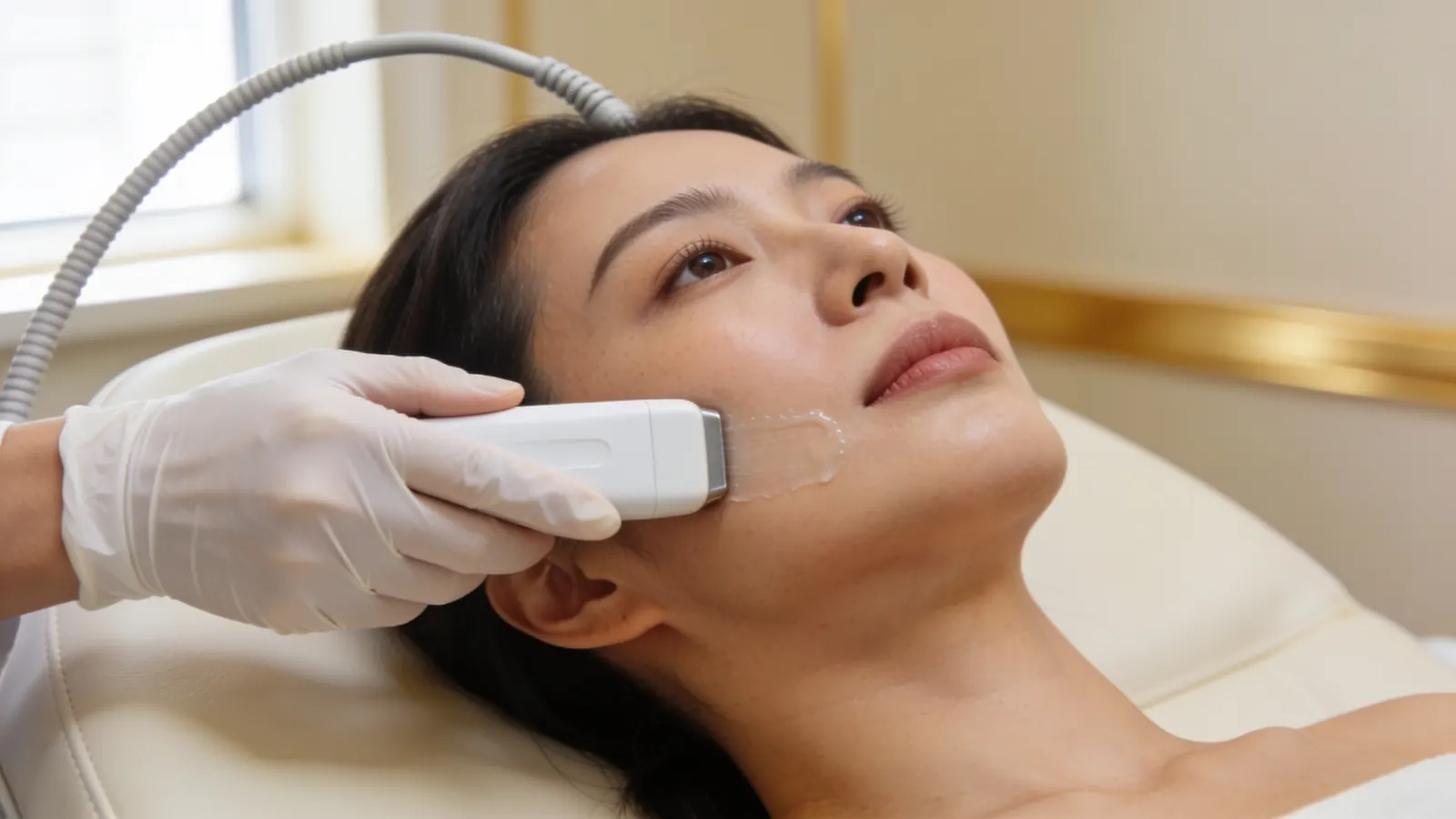
Illustrative image — not a patient.
Surgical teams image first too. One group used MRI for most patients, and ultrasound for the lips, before operating (Pascali 2023), and another sorted granulomas on imaging into cystic and infiltrating types that were treated differently (Choi 2022).
The scan also points to the treatment. In 231 patients with firm lumps after filler, ultrasound agreed with the tissue diagnosis in 85.7% of the 70 who had a biopsy, and the lumps fell into seven ultrasound types, most often an accumulation of filler that was not hyaluronic acid (37.2%) or a granuloma around such filler (22.5%). Granulomas around remaining non-hyaluronic acid filler responded better to non-invasive treatment. Granulomas with no filler left, and hyaluronic acid with a granuloma, did better with invasive treatment and invasive treatment gave greater improvement overall (Wang 2026). What the lump is on the scan, not simply that it is hard, is what points the way.
The scan helps answer the questions that decide everything else. Is it hyaluronic acid that can be dissolved? Is it a defined lump, or material spread through the tissue? Is there fluid or pus? Is it filler at all, or a cyst, a lymph node or something else? The scan shows what a lump is likely made of; it does not prove it, and where it does not look like filler, further tests are arranged. Dr Sin Yong performs the scan himself, and the plan follows what it shows. More on scanning is in ultrasound-guided filler correction.
What Dr Sin Yong does instead of cutting filler out
Maps. Dr Sin Yong scans each lump himself with ultrasound: the likely material, its depth, how far it has spread and the vessels nearby.
Treats inflammation or infection first. A red, hot, tender or draining lump needs a doctor the same day, or A&E if no doctor can see you today, and is treated medically before anything else.
Dissolves hyaluronic acid. He uses hyaluronidase, which acts on hyaluronic acid only. Your allergy history is checked. Before planned dissolving, Dr Sin Yong always does a skin test: a small amount of hyaluronidase is injected just under the skin to check for a reaction. A small first dose goes into the mapped excess, with a review at about 48 hours, because dissolving more than the excess can leave a hollow. In an emergency such as a blocked blood vessel, dissolving is not delayed for a skin test.
Treats what cannot be dissolved, without an incision. He treats silicone, polyacrylamide gel, PMMA, Ellansé, Sculptra, Juvelook, Radiesse, layered products and excess fat graft with the Sculpting mode of Time Freeze Laser LCLR®, his own protocol on a long-pulsed 755 nm and 1064 nm laser platform, directed at the mapped lump, without an incision. Like any laser, it can cause blistering or a change in skin colour, so settings are chosen for your skin.
Reviews. Each lump is reassessed, and any further visit is planned from what is seen.
Red, hot, swollen or draining: medical treatment first
A lump that is red, hot, tender, swollen or draining may be infected or actively inflamed. It needs a doctor the same day, or A&E if no doctor can see you today, and is treated medically first, before any device and before any talk of cutting it out. A collection of pus can need draining through a small opening alongside medical treatment; that is a smaller procedure than an operation to cut filler out. A fever needs a doctor the same day; if no doctor can see you today, A&E. Feeling faint, confused or very unwell: call 995 or go to A&E now.
Urgent: a change in vision or eye pain after filler: call 995 now. Skin that turns pale, dusky or mottled, or pain that is getting worse, needs urgent assessment by the doctor who injected you, or at A&E, now. See the warning signs of a blocked blood vessel after filler.
Before you agree to surgery: a checklist
Questions to ask
Has the lump been scanned, and what does the scan show it is likely made of?
Is any of it hyaluronic acid that could be dissolved first?
Is it a defined lump, or material spread through the tissue? How much do you expect to take out, and what happens to what is left?
Where will the incision be, and where will the scar sit?
Which nerves run near it, and what would an injury to them mean for feeling or movement?
Will tissue be removed with the filler? Could the area be left with a dent, and how would that be dealt with?
How likely is a second operation?
Has any inflammation or infection been treated first?
What happens if I wait, and what can be done without an incision?
Records to bring
Product names, dates, areas and amounts injected, if known, including treatments done overseas. Having no records is fine.
Photos from before the filler and from when the lump appeared.
Any scan reports or images, operation notes and tissue (histology) reports.
What has been tried, such as dissolving, medical treatment or laser, and what happened.
Your allergy history, including any reaction to hyaluronidase.
What you were offered and what you were told, in writing if you have it.
Why a scan comes first
An operation can still be done later if it is ever needed. It cannot be undone. A scan first means the decision, whichever way it goes, is made on what is actually there. If you have already been offered surgery, Dr Sin Yong can scan the lump and give you a second opinion before you decide.
Tried other treatments and almost given up?
Many patients who come to see Dr Sin Yong about filler they were told has to be cut out tell him they have tried almost everything else and had given up hope. Having tried other treatments does not mean nothing more can be done: what is done next depends on what the scan shows.
That history is useful. Knowing what was injected, when, and what has already been tried often explains why a problem has stayed: hyaluronidase acts only on hyaluronic acid and only where it reaches, some fillers are not hyaluronic acid at all, and swelling or fluid can look like filler. Ultrasound helps show which of these applies, and the plan starts from there. For material that cannot be dissolved, Dr Sin Yong uses the Sculpting mode of Time Freeze Laser LCLR® for nodules large and small, without an incision. How each person responds varies.
If you have been told nothing more can be done, it is still worth having the area scanned: ultrasound helps show what is there, and the plan is made from that. Bring any records of past treatments, product names and dates if you have them. More on this is in when filler dissolving has not worked.
How Dr Sin Yong treats filler lumps without surgery
Dr Sin Yong developed the Time Freeze Laser LCLR® protocol himself and performs every scan and treatment himself on Orchard Road. He has been invited to share his clinical expertise in Seoul, Dubai, Bangkok and São Paulo, including as a Global Key Opinion Leader.
He explains the risks and limits of each option at the assessment, including waiting where that is reasonable and surgery where it is needed. Get assessed before you agree to an operation: it can still be done later if it is ever needed, but it cannot be undone. More on the laser is in the Sculpting mode of Time Freeze Laser LCLR®.
Your next step
Not for emergencies. A change in vision or eye pain after filler: call 995 now. Pale, dusky or mottled skin, or pain that is getting worse: go now to the doctor who injected you, or to A&E. Do not message or book for these signs.
1. MessageWhatsApp +65 8023 7170 with what is bothering you. A reply comes from the clinic, not a bot.
2. AssessmentDr Sin Yong examines you himself on Orchard Road, with ultrasound where earlier filler is involved.
3. A plan in writingWhat he would do, what he would not, the fee factors, and the option of no treatment.
Every treatment is performed by Dr Sin Yong personally. Singapore Medical Council registered · invited to share his clinical expertise internationally for CLASSYS, DEKA, Fotona, Hironic and Ilooda.
Usually not. Get assessed before you agree to it. Hyaluronic acid is dissolved. Silicone, Ellansé, Sculptra, Juvelook, Radiesse, PMMA, gels and fat graft cannot be dissolved, and Dr Sin Yong treats them with the Sculpting mode of Time Freeze Laser LCLR®, without an incision. In his practice, almost every hard lump is treated without surgery. If yours is one of the few that needs surgery, you are told plainly, and why.
Many of the patients Dr Sin Yong sees have tried other treatments first. A treatment that did not help can still leave options open: hyaluronidase may not have reached the filler, the filler may not be hyaluronic acid, or what looks like filler may be swelling or fluid. Ultrasound helps show which applies, and the plan follows what it finds.
Hyaluronic acid can: it is dissolved with hyaluronidase after ultrasound mapping and a skin test. Material that cannot be dissolved is a different question. Dr Sin Yong treats it with the Sculpting mode of Time Freeze Laser LCLR®, without an incision, aiming at the lump that troubles you rather than cutting tissue out. Published work also describes drainage, and lasers placed inside the lump followed by suction, used before open surgery (Choi 2022).
A scar, because every incision leaves one; a dent or uneven contour where tissue is removed; and injury to the nerves that give the face feeling and movement. After filler was drained and a facelift done in 50 women, 4 had a facial nerve branch stop working for a time and 5 had thick, raised scars (Gadallah 2023). Bleeding under the skin (Ghareeb 2022), material left behind and repeat operations (Choi 2022) are also reported. Surgery cannot be undone.
It can be cut out, and in 35 patients with silicone granulomas of the chin the operation improved chin shape and redness. But the authors state that complete surgical removal of injected silicone is nearly impossible, because it migrates, and the granulomas returned in 44.4% after surgery alone (Harlim 2026). In Dr Sin Yong's practice, almost every silicone lump is treated without surgery: he maps it with ultrasound and treats it with the Sculpting mode of Time Freeze Laser LCLR®, without an incision.
Nodules from Ellansé (polycaprolactone) or Sculptra (poly-L-lactic acid) can be cut out, with the same risks as any filler surgery: a scar, a possible dent, nerve injury and material left behind. In a published case of granulomas in both lower eyelids after poly-D,L-lactic acid, a related collagen stimulator, two operations were needed (Garlaschi 2026). Neither product dissolves with hyaluronidase. Dr Sin Yong maps the nodules with ultrasound and treats them with the Sculpting mode of Time Freeze Laser LCLR®, without an incision.
Not always. Authors who operated on silicone granulomas of the chin state that complete surgical removal of injected silicone is nearly impossible because it migrates (Harlim 2026). After lip reconstruction for polyacrylamide gel, gel left in the lining of the lip had to be accepted (Kastner 2018), and in 35 women treated for granulomas, 4 had incomplete removal or an uneven contour (Choi 2022). Filler spread through tissue is the hardest to take out.
Whoever you see, have the lump mapped first, so the decision is informed. Ultrasound shows what the lump is likely made of, how deep it sits and how far it has spread, and that decides whether it can be dissolved, treated without an incision, or needs an operation. An international consensus calls ultrasound mandatory for managing filler complications (Velthuis 2025). If you have already been offered surgery, Dr Sin Yong can scan the lump and give you a second opinion before you decide.
Hyaluronic acid lip filler is dissolved, not cut out. Surgery is offered for material that cannot be dissolved, such as silicone or polyacrylamide gel. In 11 patients with gel spread through every layer of the lip, removal meant reconstructing the lip, with fat grafting in eight; ten had a satisfying outcome, one had severe tissue loss in the upper lip, and some gel had to be left behind (Kastner 2018). Get assessed before you agree to it.
It depends on what the scan shows. Cystic granulomas have been drained, and infiltrating ones treated with a laser fibre and suction, with open surgery only where that was not enough (Choi 2022). In 231 patients, granulomas around remaining non-hyaluronic acid filler did better with non-invasive treatment, and those with no filler left did better with invasive treatment (Wang 2026). That study did not use the Sculpting mode, and its figures do not apply to it. Dr Sin Yong treats inflammation medically first, maps the lump, dissolves any hyaluronic acid and treats other material with the Sculpting mode of Time Freeze Laser LCLR®, without an incision.
For hyaluronic acid, no: it can be dissolved. For non-absorbable filler, a group that operated on 231 patients states that surgical excision is the only way to take it out (Pascali 2023). Taking every trace out and treating the lump that troubles you are different aims, and cutting brings a scar, possible dents and nerve injury. In Dr Sin Yong's practice, almost every hard lump is treated without surgery. Get a scan and a second opinion before you decide.
If it is hyaluronic acid, it is dissolved rather than cut out: in 61 patients with long-lasting lower-eyelid swelling after hyaluronic acid filler, one hyaluronidase treatment was effective in 92% (Skippen 2020). Fat graft and collagen stimulators cannot be dissolved, and surgery for them in the lower eyelid has left dents and needed second operations (Li 2024; Garlaschi 2026). Dr Sin Yong maps under-eye material with ultrasound and treats what cannot be dissolved with the Sculpting mode of Time Freeze Laser LCLR®, without an incision.
A red, hot, painful, swollen or draining lump, a lump that is growing quickly, or a fever needs a doctor the same day, not a planned laser visit or an operation; if no doctor can see you today, go to A&E. Spreading redness with a fever, feeling faint, confused or very unwell, or swelling around the eye with pain: call 995 or go to A&E now. A change in vision or eye pain after filler is an emergency: call 995 now. Skin that turns pale, dusky or mottled, or pain that is getting worse, needs urgent assessment by the doctor who injected you, or at A&E, now.
Dr Sin Yong performs the ultrasound scan, the dissolving and every Sculpting mode treatment himself, on Orchard Road. It is not delegated to another doctor, a nurse or a therapist, so the same doctor follows each lump from the first scan to the last review. Bring any records of what was injected and what you were offered; having none is fine.
References
Facial Contouring after Permanent Filler Removal with Combined Facelift and Fat Grafting: A Case Series. Plastic and Reconstructive Surgery Global Open, 2023. source
Complicated Facial Fillers: Management Algorithm. Plastic and Reconstructive Surgery Global Open, 2022. source
Novel Granulectomy Technique and Intralesional Triamcinolone for Liquid Injectable Silicone-Induced Chin Granulomas: A Long-Term Ambidirectional Cohort Study. Journal of Clinical Medicine, 2026. source
Minimally invasive removal of facial foreign body granulomas. Archives of Aesthetic Plastic Surgery, 2022. source
Removal of Polyacrylamide Gel (Aquamid®) from the Lip as a Solution for Late-Onset Complications: Our 8-Year Experience. Aesthetic Plastic Surgery, 2018. source
Surgical Removal of Fat Nodules Formed in the Tear Trough After Autologous Fat Grafting. Aesthetic Plastic Surgery, 2024. source
Clinical outcome in a series of 173 cases of foreign body granuloma: improved outcomes with a novel surgical technique. Journal of Plastic, Reconstructive & Aesthetic Surgery, 2012. source
Complications and Management of Polymethylmethacrylate (PMMA) Injections to the Midface. Aesthetic Surgery Journal, 2016. source
Complex Surgical Management of PDLLA-Induced Lower Eyelid Granulomas. Journal of Craniofacial Surgery, 2026. source
Lower lid deformity secondary to autogenous fat transfer: a cautionary tale. Aesthetic Plastic Surgery, 2008. source
Rehabilitation of the Dysmorphic Lower Eyelid From Hyaluronic Acid Filler: What to Do After a Good Periocular Treatment Goes Bad. Aesthetic Surgery Journal, 2020. source
The use of ultrasound imaging in aesthetic injectables: A modified Delphi consensus. Journal of Plastic, Reconstructive & Aesthetic Surgery, 2025. source
Retrospective Study on Palpable Indurations After Cosmetic Filler Injection: Ultrasonic Characterization and Image-Guided Therapy. Aesthetic Surgery Journal, 2026. source
About this page: written and medically reviewed by Dr Sin Yong, MBBS (NUS), MRCS (Edinburgh) · Last reviewed 26 October 2026 · Illustrative images only, not patients · Editorial policy
Not for emergencies. A change in vision or eye pain after filler: call 995 now. Pale, dusky or mottled skin, or pain that is getting worse: go now to the doctor who injected you, or to A&E. Do not message or book for these signs.