Swelling months later that is a reaction rather than an infection: see delayed filler reaction.
Published 26 October 2026 · Reviewed by Dr Sin Yong
A red, hot, painful or swollen area after filler, pus or a fever can mean infection, soon after the injection or months to years later. Like a blocked blood vessel or a serious allergic reaction, infection is a filler problem where acting quickly matters. Have it seen by a doctor the same day; if no doctor can see you today, go to A&E. It is treated medically first, before any device and before any talk of surgery.
Short answerInfection after filler shows as redness, heat, pain and swelling, sometimes with pus or a fever, soon after the injection or months to years later. Have it seen by a doctor the same day; if no doctor can see you today, go to A&E. Spreading redness with a fever means calling 995 or going to A&E now, and difficulty breathing means calling 995 now. An expert consensus lists a dormant biofilm, bacteria living on the filler, among the proposed causes of late reactions to hyaluronic acid filler, and advises drainage, a culture and antibiotics for an infection rather than hyaluronidase first (Baranska-Rybak 2024). Any lump left afterwards is scanned and treated according to the material.
A red, hot, painful or swollen area after filler, pus or a fever needs a doctor the same day (A&E if no doctor can see you today); spreading redness with a fever means 995 or A&E now, and difficulty breathing means 995 now.
Years later
In a study of polyacrylamide gel, bacteria were found in 53 of 54 testable patients with reactions and in none of 24 without, in lesions up to five years after injection (Christensen 2013).
Biofilm
Hyaluronic acid, polyacrylamide gel and poly-L-lactic acid all supported bacterial biofilm in laboratory tests, and all six biopsies from long-standing granulomas showed it (Saththianathan 2017).
Triggers
In 85 patients with late problems from non-absorbable fillers, 28% began after dental procedures, more filler or other invasive treatments to the face (Kadouch 2013).
Treatment order
Drainage of any pus, a culture and antibiotics come before hyaluronidase (Baranska-Rybak 2024), and no laser or other device is used on an active infection.
Afterwards
Any lump that remains is scanned; hyaluronic acid is dissolved, and material that cannot be dissolved is treated with the Sculpting mode of Time Freeze Laser LCLR®, without an incision.
Red, hot or swollen after filler: what to do today
Infection after filler can start soon after the injection or months to years later. Like a blocked blood vessel or a serious allergic reaction, it is a filler problem where acting quickly matters. It is treated medically first, before any device and before any talk of surgery. A review of filler problems other than blocked blood vessels, infection among them, sorts them into early and delayed, and stresses recognising them by their timing and managing them early (Hong, Diagnostics 2024). Do not wait to see whether it settles.
A red, hot, painful or swollen area, a lump that is getting bigger, or pus: have it seen by a doctor the same day, by whoever can see you soonest, whether the doctor who injected you, Dr Sin Yong or your own doctor. If none of them can see you today, go to A&E.
Fever, or feeling unwell, with a red or swollen area: a doctor the same day, not a planned appointment; if no doctor can see you today, A&E.
Redness that is spreading, with a fever, or feeling faint, confused or very unwell: call 995 or go to A&E now. Any difficulty breathing: call 995 now.
A change in vision or eye pain after filler: call 995 now. Swelling around the eye with pain: call 995 or go to A&E now.
Difficulty breathing, a tight throat, swelling of the tongue or throat, or swelling that spreads quickly across the face, after any injection, including hyaluronidase, can be a serious allergic reaction: call 995 now. Itch, hives or swelling that spreads beyond the treated area: contact the clinic that injected you the same day.
Until you are seen, do not squeeze, massage or try to drain the lump yourself, and do not have more filler, or any laser or device treatment, on the area.
A blocked blood vessel can be mistaken for infection, because the area can be red and painful too. Skin that turns pale, dusky or mottled is a warning sign of a blocked vessel: that, or pain that keeps getting worse, needs urgent assessment by the doctor who injected you, or at A&E, now, whichever the cause.
Urgent: a change in vision or eye pain after filler: call 995 now. Skin that turns pale, dusky or mottled, or pain that is getting worse, needs urgent assessment by the doctor who injected you, or at A&E, now. See the warning signs of a blocked blood vessel after filler.
Skin that is dying after a blocked blood vessel (necrosis) is an emergency. It needs urgent, more intensive care than a lump does, starting now.
Filler infection symptoms: what an infected filler looks like
The signs of infection after filler are those of infection anywhere in the skin:
Redness around the treated area, sometimes spreading outwards.
Heat: the area feels warmer than the skin around it.
Pain and tenderness that build rather than ease.
Swelling or a lump that is getting bigger, or one that turns soft and tense, which can mean pus has collected (an abscess).
Pus or fluid draining from a point on the skin.
Fever, or feeling generally unwell.
Normal swelling and tenderness after filler ease steadily as the area settles. Infection goes the other way: redness, heat and pain that build, or that come back after the area had settled. Normal early swelling is described in filler swelling stages.
Infected lip filler
The lips swell readily after filler, so swelling alone does not mean infection. Redness spreading beyond the lip border, heat, throbbing pain, a lump that keeps growing, pus or a fever are signs of infection. Small clustered blisters can be a cold sore, which can flare after lip injections and needs different medical treatment. Blisters on skin that is pale, dusky or mottled can mean a blocked blood vessel: urgent assessment by the doctor who injected you, or at A&E, now. The lips are also an area where a blocked blood vessel is a real risk, so the urgent signs above apply. Other lip problems, including lumps that appear months later, are covered in lip filler gone wrong.
“Dissolving filler is not an undo button.”
Dr Sin YongOn what dissolving can and cannot do
Early infection, late infection, or a delayed reaction?
Problems after filler are grouped by when they start. International consensus on hyaluronic acid fillers calls a reaction delayed-onset when it starts more than four weeks after treatment (Philipp-Dormston 2020).
Early infection
An infection that starts soon after the injection usually comes from bacteria carried in at the time, from the skin or from anything the needle touches. In laboratory tests, repeated needle passes through a contaminated surface increased the contamination of the filler 10,000-fold (Saththianathan 2017). It shows as redness, heat, pain, swelling and sometimes pus at the injection sites.
Late infection
Infection can also appear months or years later. Bacteria can live on the surface of filler as a biofilm, a thin layer that shelters them, and stay quiet until something sets them off. An expert consensus on late reactions to hyaluronic acid filler lists infection introduced at the injection, or a dormant biofilm, among the proposed causes, and recent dental treatment among the risk factors (Baranska-Rybak 2024). In 85 patients with late problems from non-absorbable fillers, symptoms began after dental procedures, more filler injections or other invasive treatments to the face in 28% (Kadouch 2013).
Not every late red lump is infection
A lump that flares months later can also be the immune system reacting to the filler: a delayed inflammatory reaction. These are uncommon, about 1.1% a year in prospective studies of hyaluronic acid filler (Chung 2020). After 4,702 treatments with one hyaluronic acid filler, 23 patients developed delayed-onset nodules, a median of four months after injection; 9 of them had an identifiable immune trigger such as a flu-like illness, and the authors consider the nodules likely immune-mediated (Beleznay 2015).
From the outside, the two can look alike. Either way, a red, inflamed lump needs a doctor the same day (A&E if no doctor can see you today) and is treated medically first. Examination, ultrasound and, where there is pus, a culture help tell them apart, and that decides what comes next. Delayed reactions are covered in delayed filler reaction, and reactions that form a firm lump around the product in filler granuloma.
Can filler get infected years later? Biofilm after filler
Yes. Filler can stay in the tissue for years, and bacteria that reach it can stay with it. These are the findings behind that answer:
Biofilm forms on filler. In laboratory tests, hyaluronic acid, polyacrylamide gel and poly-L-lactic acid all supported bacterial biofilm, and all six biopsies from five patients with long-standing granulomas after filler showed bacterial biofilm (Saththianathan 2017).
Bacteria in gel lumps, years after injection. In a study of polyacrylamide gel, bacteria, mostly ordinary skin bacteria in small clusters, were found in 53 of the 54 patients with reactions who could be tested and in none of 24 without reactions, in lesions up to five years after injection. The authors conclude that low-grade bacteria can cause long-term infection with the gel, possibly as a biofilm (Christensen 2013).
Infections years after treatment. In 267 people treated with polyalkylimide gel (Bio-Alcamid) for facial fat loss related to HIV, infections were documented in 56, a median of 32 months after the first treatment; touch-up injections, cosmetic surgery, facial injury and dental work were among the possible risk factors (Nadarajah 2012).
An abscess that kept coming back. In a published case, an abscess under the chin kept returning after hyaluronic acid filler to the chin; the authors weighed bacterial contamination against an immune reaction (Nicola 2024).
Much of this evidence comes from gels that the body does not absorb, but laboratory work shows that hyaluronic acid can carry a biofilm too (Saththianathan 2017). A lump that keeps flaring is therefore assessed for infection before anything else is done to it. Lumps from non-absorbable gels have their own pages: polyacrylamide filler lumps and silicone and other non-absorbable filler lumps.
Infected filler by type
Swipe sideways to see the whole table.
Filler
Can it be dissolved?
What is reported about infection
How Dr Sin Yong approaches it
Hyaluronic acid (most lip, cheek, chin and under-eye fillers)
Yes, with hyaluronidase
Supported bacterial biofilm in laboratory tests (Saththianathan 2017); an abscess under the chin that kept returning (Nicola 2024); a rare mycobacterial infection (Liu, Clin Cosmet Investig Dermatol 2023)
Infection treated medically first, with drainage of any pus; the filler is then mapped and dissolved, after a skin test
Polyacrylamide gel (Aquamid and similar gels)
No
Bacteria in 53 of 54 testable patients with reactions, in lesions up to five years after injection (Christensen 2013); supported biofilm in laboratory tests (Saththianathan 2017)
Infection treated medically first, with drainage of any pus; what remains is mapped and treated with the Sculpting mode of Time Freeze Laser LCLR®, without an incision
Polyalkylimide (Bio-Alcamid)
No
In people treated for HIV-related facial fat loss, infections a median of 32 months after the first treatment; most needed surgical drainage as well as antibiotics (Nadarajah 2012)
Infection treated medically first, with drainage of any pus; what remains is mapped and treated with the Sculpting mode, without an incision
Silicone, PMMA and other non-absorbable fillers
No
In 85 patients with late problems from non-absorbable fillers, 28% began after dental procedures, more filler or other invasive facial treatments; abscesses were more frequent in people with HIV-related facial fat loss (Kadouch 2013)
Infection treated medically first; what remains is mapped and treated with the Sculpting mode, without an incision
Poly-L-lactic acid, the material in Sculptra, supported bacterial biofilm in laboratory tests (Saththianathan 2017)
Infection treated medically first; nodules are then mapped and treated with the Sculpting mode, without an incision
Mesotherapy and other skin injections (not filler)
Only if it is hyaluronic acid
Rapidly growing mycobacteria in 16 patients, all of whom needed surgical drainage (Regnier 2009)
Samples tested for mycobacteria; drainage, surgical where needed, with specialist infection care; a lump that remains once healed is scanned and treated according to the material
Unknown or mixed material
Only any hyaluronic acid part
Often injected years ago or overseas, without records
Infection treated first; then scanned; hyaluronic acid dissolved; what remains treated with the Sculpting mode, without an incision
Unusual infections after injections: mycobacteria
Rarely, an infection after an injection is caused by a mycobacterium, one of a group of bacteria related to the one that causes tuberculosis.
After hyaluronic acid filler. A published case describes a skin infection with Mycobacterium abscessus after hyaluronic acid was injected into the face. It was identified with laboratory tests and treated by drainage and surgical removal of the affected tissue (Liu, Clin Cosmet Investig Dermatol 2023).
After mesotherapy. In a series of 16 patients who had mesotherapy, skin injections of other substances rather than filler, painful, red, draining lumps appeared at the injection sites, caused by rapidly growing mycobacteria, mostly M. chelonae. All needed surgical drainage, antibiotics were given for an average of 14 weeks, and healing took an average of 6.2 months (Regnier 2009).
The practical point: a lump that keeps draining, or does not settle with the usual treatment, needs a sample of pus or tissue sent to the laboratory with a request to look for mycobacteria, because they are treated differently. They are one more reason a red, draining lump after any injection is seen by a doctor the same day, or at A&E if no doctor can see you today, rather than watched.
How a filler infection is treated: medical treatment first
An infected filler lump is treated medically first, before any device and before any talk of surgery. This is what that involves:
Antibiotics. Chosen for the likely bacteria and changed if the laboratory report points elsewhere. For late nodules after hyaluronic acid filler, an international panel listed antibiotics by mouth or injected into the nodule among the treatments, alongside other medical treatment, hyaluronidase for nodules that are not inflamed, and surgical excision as a last resort (Philipp-Dormston 2020).
Drainage of any pus. A collection of pus is let out through a small opening, with a needle or a small nick in the skin, alongside the antibiotics. This is a much smaller procedure than an operation to cut filler out.
A culture. A sample of the pus is taken, where possible before antibiotics start and without delaying them, and sent to the laboratory to identify the bacteria and the antibiotics that act on them. An expert consensus on late reactions to hyaluronic acid filler advises drainage, a culture and antibiotics for an infection, rather than hyaluronidase as the first step (Baranska-Rybak 2024).
Close review. The area is checked again to make sure it is settling. Redness that spreads, a new fever or feeling worse means a doctor the same day (A&E if no doctor can see you today); spreading redness with a fever, or feeling very unwell, means calling 995 or going to A&E now.
Can infected hyaluronic acid filler be dissolved?
Hyaluronic acid can be dissolved with hyaluronidase, but for an infection that is not the first step (Baranska-Rybak 2024). Once the infection is under control on antibiotics and any pus has been drained, the hyaluronic acid that remains can be dissolved, because the filler is where bacteria can shelter as a biofilm. In the published case of an abscess under the chin that kept returning after hyaluronic acid filler, the authors managed it with ultrasound-guided hyaluronidase (Nicola 2024). That is a single case. In Dr Sin Yong's practice, dissolving never replaces treating the infection. How dissolving is done is set out below.
Infection in filler that cannot be dissolved
Polyacrylamide gel, polyalkylimide, silicone, PMMA, Sculptra, Ellansé, Radiesse and the poly-D,L-lactic acid in Juvelook do not dissolve. Infection in or around them is treated with antibiotics and drainage of any pus; in the polyalkylimide series above, most infections needed surgical drainage as well as antibiotics (Nadarajah 2012). Once the infection has settled, the material that remains can still be treated without an incision: Dr Sin Yong treats it with the Sculpting mode of Time Freeze Laser LCLR®.
Why lasers and other devices are not used on an active infection
Dr Sin Yong does not use the Sculpting mode of Time Freeze Laser LCLR®, or any other laser or energy device, on a lump that is actively infected or inflamed. The reasons are practical:
It does not replace medical treatment. An infection needs medical treatment, and pus needs a way out. A treatment given without an incision does not drain it.
Time matters. A visit spent on a device is time in which the infection is not being treated.
Swelling hides what is underneath. While the area is inflamed, part of what can be felt is swelling, not filler. Once the infection has settled, the area is scanned again to see what, if anything, remains.
The same applies to massage, radiofrequency and other energy treatments, and to more filler: none of them is used on an infected area, and a blocked blood vessel is never treated with the Sculpting mode or any other laser. Once it has settled, a lump that remains is treated according to what it is made of.
After the infection has settled: scanning and treating what remains
Some infected lumps settle completely with treatment. Others leave filler, scar tissue or a firm lump behind. Once the infection has settled, the area is scanned again.
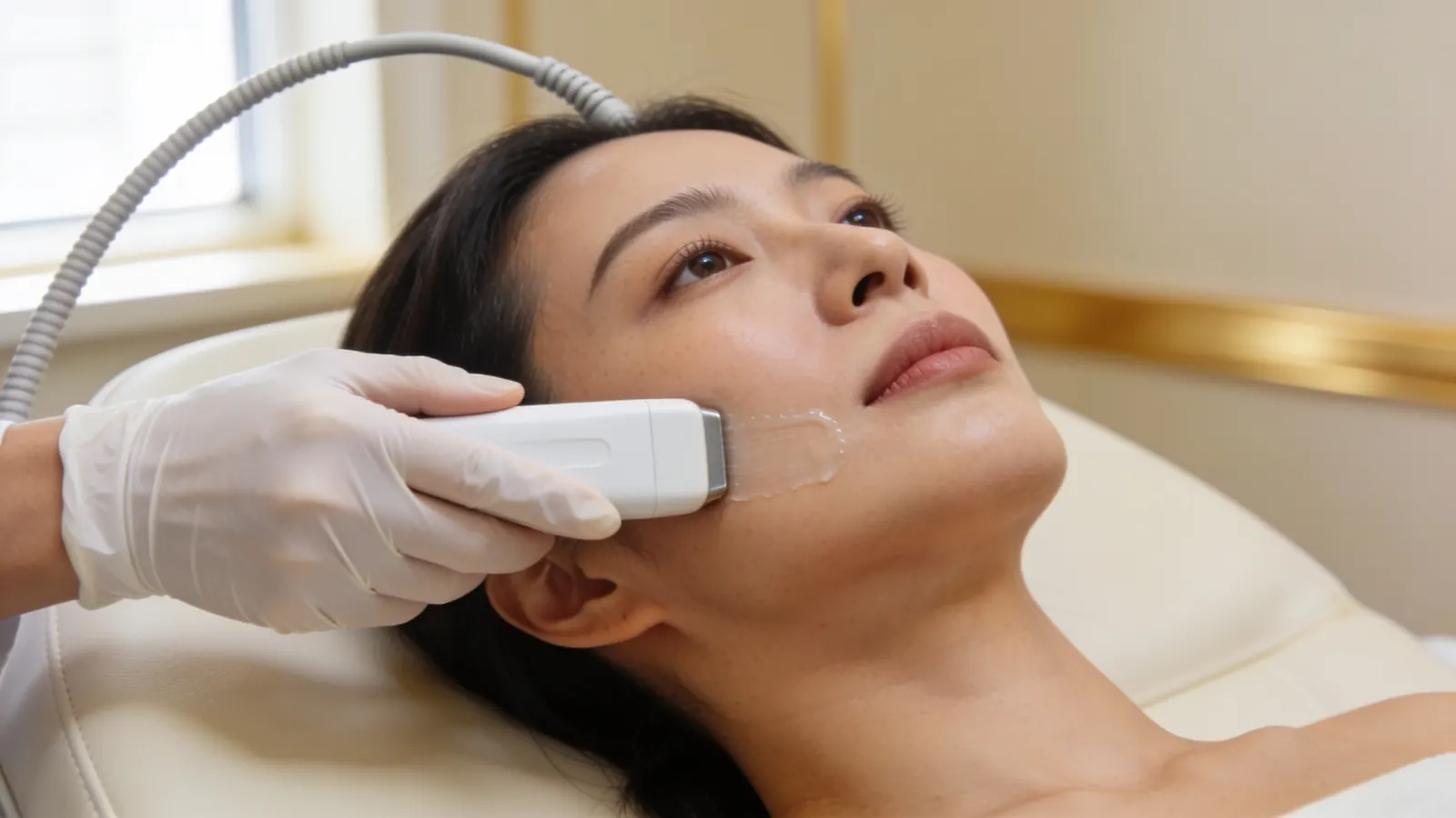
Illustrative image — not a patient.
Ultrasound shows where the filler sits, how deep it is and how far it has spread, and what it is likely made of. It also shows whether a collection of fluid, such as pus, is still there. An international consensus of 15 experts from seven specialties calls ultrasound mandatory for managing filler complications, and states that ultrasound guidance is needed for precise injections into a lesion (Velthuis 2025). In a study of 191 people with filler around the eyes, a blood-flow signal within an area of filler on the scan suggested infection (Zhang 2025). The scan shows what a lump is likely made of; it does not prove it, and where it does not look like filler, further tests are arranged. Dr Sin Yong performs the scan himself. More on scanning is in ultrasound-guided filler correction.
Hyaluronic acid: dissolved
Hyaluronic acid that remains is dissolved with hyaluronidase, which acts on hyaluronic acid only. Your allergy history is checked. Before planned dissolving, Dr Sin Yong always does a skin test: a small amount of hyaluronidase is injected just under the skin to check for a reaction. A small first dose goes into the filler mapped on the scan, with a review at about 48 hours, because dissolving more than the excess can leave a hollow. In an emergency such as a blocked blood vessel, dissolving is not delayed for a skin test. Dr Sin Yong waits at least two weeks after hyaluronidase before any refilling. More is in hyaluronidase and hyaluronidase allergy and the skin test.
Material that cannot be dissolved: treated without an incision
Silicone, polyacrylamide gel, polyalkylimide, PMMA, Sculptra, Ellansé, Radiesse, the poly-D,L-lactic acid in Juvelook and excess fat graft cannot be dissolved, but they can still be treated without an incision. Dr Sin Yong maps the lump and treats it with the Sculpting mode of Time Freeze Laser LCLR®, his own protocol on a long-pulsed 755 nm and 1064 nm laser platform, directed at the mapped lump. Like any laser, it can cause blistering or a change in skin colour, so settings are chosen for your skin. Where hyaluronic acid sits with other material, dissolving and the Sculpting mode are planned in order. More is in the Sculpting mode of Time Freeze Laser LCLR®.
Infected filler lump, and only offered surgery? Get assessed first
Draining pus and cutting filler out are different things. Drainage lets pus out through a small opening, alongside medical treatment. An operation to remove infected filler means cutting out the lump and the tissue around it. Before you agree to that, know what it means:
A scar. Every cut through the skin of the face heals with a scar.
A dent. Cutting out a lump takes tissue with it, and the area can heal hollow or uneven.
Nerve injury. Nerves that give the face feeling and movement run close to where filler sits. An injured nerve can leave numbness, or weakness in a smile, a lip or a brow.
Product left behind. Filler spread through tissue is hard to remove completely.
No way back. Tissue that has been cut out cannot be put back.
An incision is also a new wound that can itself become infected. After non-absorbable filler was drained and a facelift with fat grafting done in 50 women, 2 had wound infections, 5 had thick, raised scars and 4 had a branch of the facial nerve that moves the lower lip stop working for a time (Gadallah 2023).
Surgery has a place. Every patient in the mesotherapy series needed surgical drainage (Regnier 2009), the mycobacterial infection after hyaluronic acid was treated by drainage and surgical removal of the affected tissue (Liu, Clin Cosmet Investig Dermatol 2023), and most infections after polyalkylimide gel needed surgical drainage as well as antibiotics (Nadarajah 2012). International consensus on delayed reactions to hyaluronic acid filler lists surgical excision as a last resort for late nodules (Philipp-Dormston 2020). The point is the order: the infection is treated and any pus drained first, the area is scanned once it has settled, and any decision to operate comes after that.
Once the infection has been treated, a lump that remains is treated like any other hard lump: in Dr Sin Yong's practice, almost every hard lump is treated without surgery. If yours is one of the few that needs surgery, you are told plainly, and why. If you have already been offered an operation, Dr Sin Yong can scan the area and give you a second opinion before you decide. What removal surgery involves is set out in filler removal surgery.
Tried other treatments and almost given up?
Many patients who come to see Dr Sin Yong about lumps left after a filler infection tell him they have tried almost everything else and had given up hope. Having tried other treatments does not mean nothing more can be done: what is done next depends on what the scan shows.
That history is useful. Knowing what was injected, when, and what has already been tried often explains why a problem has stayed: hyaluronidase acts only on hyaluronic acid and only where it reaches, some fillers are not hyaluronic acid at all, and swelling or fluid can look like filler. Ultrasound helps show which of these applies, and the plan starts from there. For material that cannot be dissolved, Dr Sin Yong uses the Sculpting mode of Time Freeze Laser LCLR® for nodules large and small, without an incision. How each person responds varies.
If you have been told nothing more can be done, it is still worth having the area scanned: ultrasound helps show what is there, and the plan is made from that. Bring any records of past treatments, product names and dates if you have them. More on this is in when filler dissolving has not worked.
How Dr Sin Yong approaches a filler infection
Dr Sin Yong examines the area and performs the ultrasound scan himself on Orchard Road. A red, hot, tender, swollen or draining lump needs a doctor the same day, or A&E if no doctor can see you today, and is treated medically first; nothing is dissolved, lasered or cut out until the infection is under control. Where a lump remains once it has settled, he maps it, dissolves hyaluronic acid and treats material that cannot be dissolved with the Sculpting mode of Time Freeze Laser LCLR®, without an incision.
He developed the Time Freeze Laser LCLR® protocol himself and has been invited to share his clinical expertise in Seoul, Dubai, Bangkok and São Paulo, including as a Global Key Opinion Leader. You do not need to have been treated here. Bring any records of what was injected, where and when; having none is fine. Lumps from every kind of filler are covered in hard lumps after filler, and other problems in dermal filler gone wrong.
Your next step
Not for emergencies. A change in vision or eye pain after filler: call 995 now. Pale, dusky or mottled skin, or pain that is getting worse: go now to the doctor who injected you, or to A&E. Do not message or book for these signs.
1. MessageWhatsApp +65 8023 7170 with what is bothering you. A reply comes from the clinic, not a bot.
2. AssessmentDr Sin Yong examines you himself on Orchard Road, with ultrasound where earlier filler is involved.
3. A plan in writingWhat he would do, what he would not, the fee factors, and the option of no treatment.
Every treatment is performed by Dr Sin Yong personally. Singapore Medical Council registered · invited to share his clinical expertise internationally for CLASSYS, DEKA, Fotona, Hironic and Ilooda.
Redness, heat, pain and swelling around the filler that build rather than ease, a lump that is growing, pus or fluid draining from the skin, and sometimes a fever or feeling unwell. It can start soon after the injection or months to years later. Normal swelling after filler eases steadily; infection builds. Have it seen by a doctor the same day; if no doctor can see you today, go to A&E. Call 995 or go to A&E now for spreading redness with a fever, and call 995 now for any difficulty breathing.
Many of the patients Dr Sin Yong sees have tried other treatments first. A treatment that did not help can still leave options open: hyaluronidase may not have reached the filler, the filler may not be hyaluronic acid, or what looks like filler may be swelling or fluid. Ultrasound helps show which applies, and the plan follows what it finds.
It needs a doctor the same day; if no doctor can see you today, A&E. A red, hot, swollen or painful lump can be infection, which is treated medically first. Spreading redness with a fever means calling 995 or going to A&E now, and any difficulty breathing means calling 995 now. A blocked blood vessel can also look red and painful. A change in vision or eye pain after filler means calling 995 now; skin that turns pale, dusky or mottled, or pain that keeps getting worse, needs urgent assessment by the doctor who injected you, or at A&E, now.
The lips swell readily after filler, so swelling alone does not mean infection. Redness spreading beyond the lip border, heat, throbbing pain, a lump that keeps growing, pus or a fever are signs of infection. Small clustered blisters can be a cold sore, which needs different medical treatment; blisters on pale, dusky or mottled skin can mean a blocked blood vessel. Infected lip filler needs a doctor the same day, or A&E if no doctor can see you today, and is treated medically first, with drainage of any pus. The lips are also an area where a blocked blood vessel is a real risk, so pale, dusky or mottled skin needs urgent assessment by the doctor who injected you, or at A&E, now.
Yes. In a study of polyacrylamide gel, bacteria were found in 53 of 54 testable patients with reactions and in none of 24 without, in lesions up to five years after injection (Christensen 2013). After polyalkylimide gel, infections appeared a median of 32 months after the first treatment in people treated for HIV-related facial fat loss (Nadarajah 2012). Hyaluronic acid also supported bacterial biofilm in laboratory tests (Saththianathan 2017). A lump that flares long after filler is assessed for infection first.
A biofilm is a community of bacteria living on a surface, here the filler, inside a protective layer they make. An expert consensus lists a dormant biofilm among the proposed causes of late reactions to hyaluronic acid filler (Baranska-Rybak 2024). In laboratory tests, hyaluronic acid, polyacrylamide gel and poly-L-lactic acid all supported biofilm, and all six biopsies from long-standing granulomas after filler showed it (Saththianathan 2017). A lump that keeps flaring is assessed for infection before anything else is done.
An abscess is a collection of pus in or around the filler: a red, hot, painful lump that can turn soft and tense, sometimes with a fever. It needs a doctor the same day, or A&E if no doctor can see you today, and is treated with antibiotics and drainage through a small opening, a much smaller procedure than cutting filler out, and the pus is sent for a culture. In a published case, an abscess under the chin kept returning after hyaluronic acid filler; the authors weighed bacterial contamination against an immune reaction (Nicola 2024).
Medically first, before any device and before any talk of surgery: antibiotics, drainage of any pus through a small opening, and a culture to identify the bacteria. An expert consensus on hyaluronic acid filler advises this rather than hyaluronidase as the first step (Baranska-Rybak 2024). Once the infection has settled, any lump that remains is scanned: hyaluronic acid is dissolved, and material that cannot be dissolved is treated with the Sculpting mode of Time Freeze Laser LCLR®, without an incision.
Yes, once the infection is under control. An expert consensus advises drainage, a culture and antibiotics for an infection, and keeps hyaluronidase for reactions without active infection (Baranska-Rybak 2024). In a published case of an abscess under the chin that kept returning after hyaluronic acid filler, which the authors discussed as bacterial contamination or an immune reaction, they managed it with ultrasound-guided hyaluronidase (Nicola 2024). Before planned dissolving, Dr Sin Yong checks your allergy history, always does a skin test first, and maps the filler with ultrasound.
Usually not. Get assessed first. An infected lump is treated medically, and pus is let out through a small opening, a much smaller procedure than cutting filler out. Some need surgical drainage: most infections after polyalkylimide gel did in one series (Nadarajah 2012), and all the mycobacterial infections after mesotherapy did in another (Regnier 2009). Cutting filler out leaves a scar, can leave a dent or injure a nerve, and cannot be undone. In Dr Sin Yong's practice, almost every hard lump is treated without surgery. If yours is one of the few that needs surgery, you are told plainly, and why.
Not while it is infected. A laser or other device does not replace medical treatment for an infection, and a treatment given without an incision does not drain pus. Dr Sin Yong treats the infection medically first and does not use the Sculpting mode of Time Freeze Laser LCLR® on an active infection. Once it has settled, the area is scanned, and material that cannot be dissolved, such as silicone or polyacrylamide gel, can then be treated with the Sculpting mode, without an incision.
Dental treatment has been linked with late problems. An expert consensus lists recent dental treatment among the risk factors for late reactions to hyaluronic acid filler (Baranska-Rybak 2024). In 85 patients with late problems from non-absorbable fillers, 28% began after dental procedures, more filler or other invasive facial treatments (Kadouch 2013), and dental work was among the possible risk factors for infections after polyalkylimide gel for HIV-related facial fat loss (Nadarajah 2012). Tell your dentist which fillers you have, and have a red, painful swelling seen by a doctor the same day, or at A&E if no doctor can see you today.
From the outside they can look alike: a red, swollen, tender lump weeks or months after filler. Delayed inflammatory reactions after hyaluronic acid filler are uncommon, about 1.1% a year in prospective studies (Chung 2020), and in one series 9 of 23 patients with delayed-onset nodules had an immune trigger such as a flu-like illness (Beleznay 2015). Either way, a red, inflamed lump needs a doctor the same day (A&E if no doctor can see you today) and is treated medically first. Examination, ultrasound and, where there is pus, a culture help tell them apart, and that decides what comes next.
Mycobacteria are bacteria related to the one that causes tuberculosis, and they are a rare cause of infection after injections. A published case describes a Mycobacterium abscessus skin infection after hyaluronic acid was injected into the face, treated by drainage and surgical removal of the affected tissue (Liu, Clin Cosmet Investig Dermatol 2023). After mesotherapy, rapidly growing mycobacteria caused painful, draining lumps in 16 patients, all of whom needed surgical drainage (Regnier 2009). A lump that keeps draining needs samples tested for them.
References
Late-Onset Reactions after Hyaluronic Acid Dermal Fillers: A Consensus Recommendation on Etiology, Prevention and Management. Dermatology and Therapy, 2024. source
Global Approaches to the Prevention and Management of Delayed-onset Adverse Reactions with Hyaluronic Acid-based Fillers. Plastic and Reconstructive Surgery Global Open, 2020. source
The Role of Bacterial Biofilm in Adverse Soft-Tissue Filler Reactions: A Combined Laboratory and Clinical Study. Plastic and Reconstructive Surgery, 2017. source
Bacterial infection as a likely cause of adverse reactions to polyacrylamide hydrogel fillers in cosmetic surgery. Clinical Infectious Diseases, 2013. source
Infectious complications of Bio-Alcamid filler used for HIV-related facial lipoatrophy. Clinical Infectious Diseases, 2012. source
Delayed-onset complications of facial soft tissue augmentation with permanent fillers in 85 patients. Dermatologic Surgery, 2013. source
Clinical Management With High-Frequency Ultrasound of Recurrent Submental Abscess Formation After Filler Placement: Bacterial Contamination or Immune-Mediated Adverse Event?. Cureus, 2024. source
Surgical Treatment for Cutaneous Mycobacterium abscessus Infection Caused by Injections of Hyaluronic Acid. Clinical, Cosmetic and Investigational Dermatology, 2023. source
Clinical management of rapidly growing mycobacterial cutaneous infections in patients after mesotherapy. Clinical Infectious Diseases, 2009. source
A Systematic Review of the Literature of Delayed Inflammatory Reactions After Hyaluronic Acid Filler Injection to Estimate the Incidence of Delayed Type Hypersensitivity Reaction. Aesthetic Surgery Journal, 2020. source
Delayed-onset nodules secondary to a smooth cohesive 20 mg/mL hyaluronic acid filler: cause and management. Dermatologic Surgery, 2015. source
Review of the Adverse Effects Associated with Dermal Filler Treatments: Part I Nodules, Granuloma, and Migration. Diagnostics, 2024. source
The use of ultrasound imaging in aesthetic injectables: A modified Delphi consensus. Journal of Plastic, Reconstructive & Aesthetic Surgery, 2025. source
Application of High-frequency Ultrasound for Detection and Characterization of Dermal Fillers in the Periorbital Region. Plastic and Reconstructive Surgery Global Open, 2025. source
Facial Contouring after Permanent Filler Removal with Combined Facelift and Fat Grafting: A Case Series. Plastic and Reconstructive Surgery Global Open, 2023. source
About this page: written and medically reviewed by Dr Sin Yong, MBBS (NUS), MRCS (Edinburgh) · Last reviewed 26 October 2026 · Illustrative images only, not patients · Editorial policy
Not for emergencies. A change in vision or eye pain after filler: call 995 now. Pale, dusky or mottled skin, or pain that is getting worse: go now to the doctor who injected you, or to A&E. Do not message or book for these signs.